
|
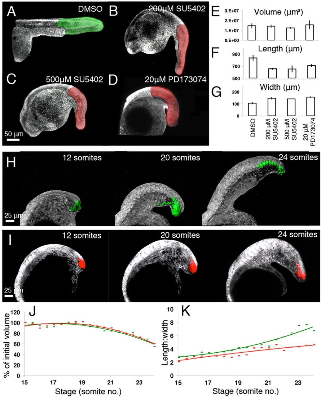
Fig. 6
FGF signalling is required for thinning and lengthening, but not volumetric growth of the zebrafish posterior body. (A-D) Embryos injected at the one-cell stage with nuclear mCherry mRNA and incubated in either 500µM DMSO (A), 200µM SU5402 (B), 500µM SU5402 (C) or 20µM PD173074 (D) from the ten-somite stage until the 32-somite stage and imaged by confocal microscopy at the 32-somite stage. (E-G) Mean value of posterior body volume (E), length (F) and width (G) for each treatment group at the 32-somite stage. Error bars indicate s.d. (H,I) Stills from time-lapse movies of embryos in which small regions within the tailbud are photolabelled at the 12-somite stage in the presence of 500µM DMSO (H) or 20µM PD173074 (I). (J,K) Quantification of the volume (J) and length:width ratio (K) of the photolabelled region over time (number of somites). Green lines correspond to the control situation (500µM DMSO), red lines indicate treatment with PD173074.