
|
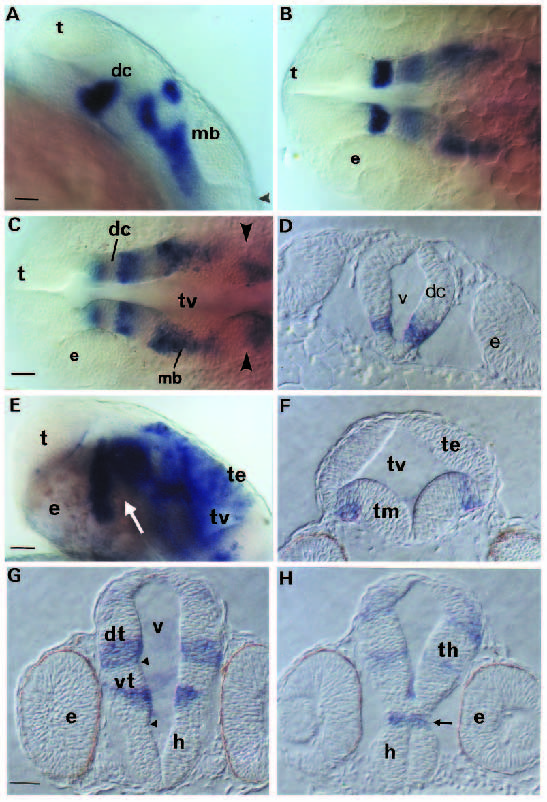
Fig. 7
Analysis of hlx-1 expression in the rostral brain. (A-C) Side view and dorsal views at two different focal planes of the rostral brain in the same 22 hpf embryo, respectively. Arrowheads mark the position of the furrow at the midbrain-hindbrain border. (D) Cross-section in the diencephalic region at 22 hpf. (E) Side view of the rostral brain of a 30 hpf embryo. Cross-sections of the midbrain (F) and diencephalic region (G,H) of a 30 hpf embryo (G is most rostral). The arrows in E and H indicate the location of the ventral flexure. Arrowheads in G mark the positions of sulci. Bar, 30 µm. Abbreviations: dc, diencephalon; dt, dorsal thalamus; e, eye; h, hypothalamus; mb, midbrain; t, telencephalon; te, tectum; th, thalamus; tm, tegmentum; tv, tectal ventricle; v, ventricle; vt, ventral thalamus.