
|
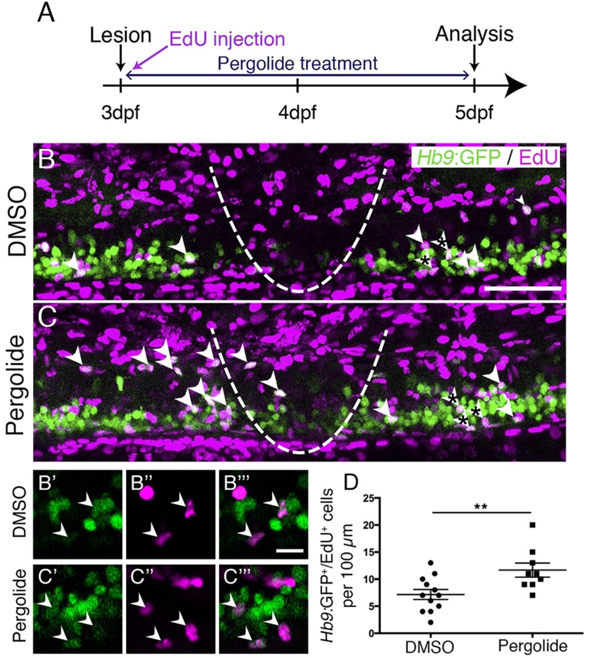
Fig. 4
Motor neuron regeneration is enhanced by application of a dopamine agonist. (A) Experimental time line. (B,C) Double-labelled Hb9:GFP+/EdU+ neurons are indicated by arrowheads. (B′-C′′′) Double-labelled cells from B,C (asterisks) are indicated by arrowheads in single optical sections at higher magnification. (D) Pergolide treatment during the regeneration phase significantly increases the number of Hb9:GFP+/EdU+ double-labelled motor neurons (t-test, **P=0.0092; DMSO, n=12; Pergolide, n=9). Lateral views are shown; rostral is left, dorsal is up. The lesion site is indicated by a dashed line. Values are means ± s.e.m. Scale bars: 50µm in B for B,C; 10µm in B′′′ for B′-C′′′.