
|
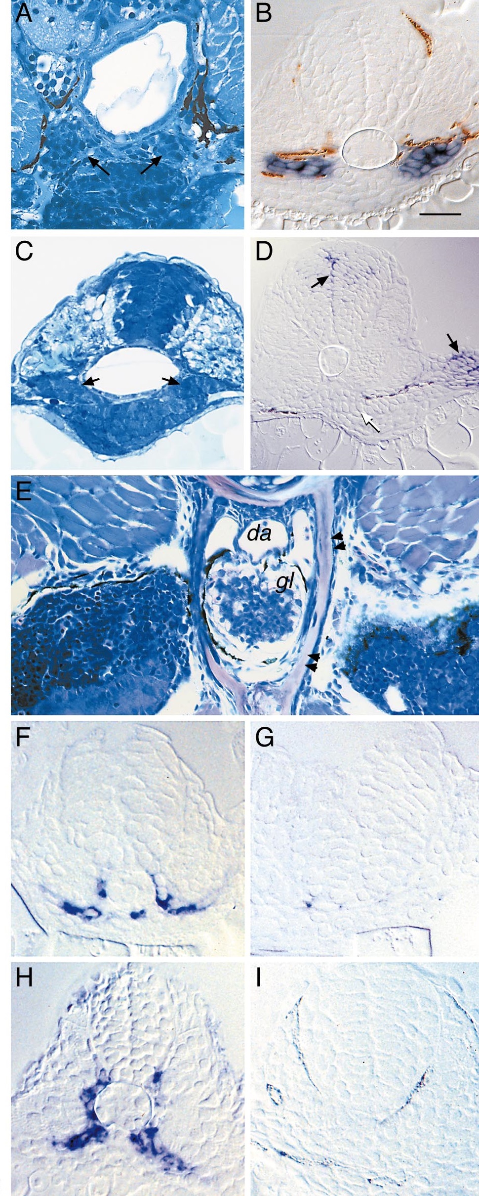
Fig. 2
Glomerular defects in sonic you and you-too mutants correlate with failed sclerotome development. Transverse section of a 48-hpf syutbx392/t4 embryo (A) reveals the absence of a midline glomerulus and in its place are two lateral cell clusters (arrows). In situ hybridization of syutbx392/t4 embryos with a wt1 probe (B) identifies the lateral cell clusters as podocytes. Similar histological section of a 36-hpf you-too embryo (C) shows a failed convergence of podocytes. In situ hybridization with gli2 probe (D) shows expression in the neural tube and fin bud (dark arrows) but no expression in the pronephric primordia (light arrow). Transverse histological section through a wildtype 3-week-old zebrafish (E) shows the glomerulus (gl) is surrounded by bone and cartilaginous tissue (double arrows; da, dorsal aorta). twist-positive cells are found in the wildtype ventromedial somite at 18 hpf (F). twist-positive cells are absent in flh mutants (G). Pax9-expressing cells are found between neural tube and somite tissue in 24-hpf wildtype embryos (H). Pax9-expressing cells are reduced in flh (I). Scale bar equals 50 µm.
Reprinted from Developmental Biology, 222(1), Majumdar, A. and Drummond, I.A., The zebrafish floating head mutant demonstrates podocytes play an important role in directing glomerular differentiation, 147-157, Copyright (2000) with permission from Elsevier. Full text @ Dev. Biol.