
|
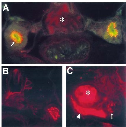
Fig. 4
flh embryos make functional glomeruli as assayed by filtration of rhodamine dextran dye. Rhodamine dextran dye (red) has been injected into the sinus venosus, filtered through the glomerulus, and taken up into duct endosomes (arrow) in 2-dpf wildtype embryos (A, B). Note that no dye is retained in the glomerulus (asterisk in A). WGA staining (green) positively identifies the duct apical brush border. In 2-dpf flh embryos (C) injected with rhodamine dextran, dye is found in the cardinal vein (arrowhead), ectopic glomeruli (asterisk), and duct endosomes (arrow). Dye is frequently trapped in glomeruli. Sections in B and C have not been stained with WGA.
Reprinted from Developmental Biology, 222(1), Majumdar, A. and Drummond, I.A., The zebrafish floating head mutant demonstrates podocytes play an important role in directing glomerular differentiation, 147-157, Copyright (2000) with permission from Elsevier. Full text @ Dev. Biol.