
|
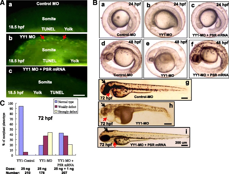
Fig. 8
Injection of PSR mRNA rescued embryos from YY1a morpholino-induced defects. YY1-MO (25 ng) and PSR mRNA (1 ng) were co-injected at the one-to-two cell stage, and embryos were assessed at 18.5 hpf, 48 hpf and 3 dpf. a Apoptotic cell analysis by TUNEL assay. Control-MO and YY1-MO (25 ng per embryo) or YY1-MO (25 ng per embryo) plus PSR mRNA (1 ng) were injected at the one-cell stage to express extra PS receptor. The embryos were fixed and observed at the different stages postfertilization (pf). TUNEL stained embryos (all at 18.5 hpf) are shown in A:a (Control-MO), A:b (YY1-MO group) and A:c (extra PSR mRNA group). The TUNEL-positive cells under the fluorescence microscope are considered apoptotic, especially in A:b (indicated by arrows). Bars indicate 100 µm. (B) Rescued embryos show normal morphology in the brain, heart, and somites. The normal morphology in the brain and heart (indicated by arrows) seen in rescued embryos (panel c, at 24 hpf; f, at 48 hpf; i, at 72 hpf) is in contrast to the deformed brains (indicated by black arrows) and hearts (indicated by red arrows) seen in YY1-MO knockdown embryos (panel b, at 24 hpf; e, at 48 hpf; h, at 72 hpf; indicated by arrows). The control-MO-injected (panel a, at 24 hpf; d, at 48 hpf; g, at 72 hpf) are the negative controls. Bars indicate 200 µm. c The ability of PSR mRNA injection to rescue embryos from morphological deformities during development at 72 hpf was estimated