
|
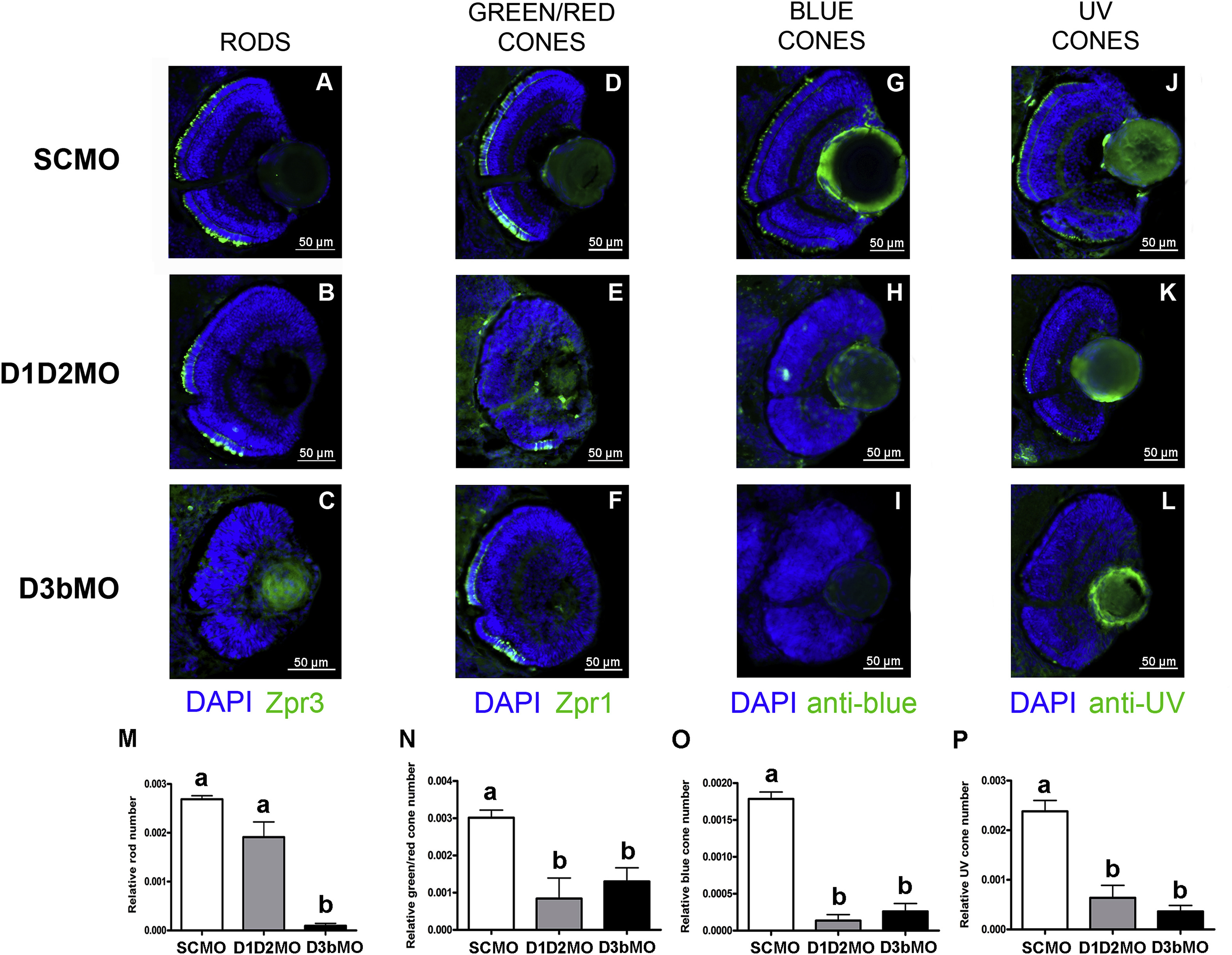
Fig. 6 Effects of deiodinase KD on relative rod and cone photoreceptor number. (A-L) Immunohistochemical stainings with Zpr3, Zpr1, anti-blue opsin and anti-UV opsin antibodies on cryosections of the eye of control (A,D,G,J), D1D2MO (B,E,H,K) and D3bMO (C,F,I,L) larvae at 3 dpf. Zpr1 is a marker for both green and red cone photoreceptors, Zpr3 is a marker for rod photoreceptors and anti-blue and anti-UV opsin label the outer segment of blue and UV cone photoreceptors respectively. Blue: DAPI staining of the nuclei; Green: Zpr1/Zpr3+Alexa488 or anti-blue/anti-UV + FITC. (M-P) Data for relative rod/cone number (expressed per retinal surface area) are shown as mean + SEM (n ≥ 5 per group). Groups with no common letter are significantly different (p < 0.05). Data in (N,O,P) followed Gaussian distribution and were analyzed by one-way ANOVA with Tukey post hoc test. Data in (M) did not assume Gaussian distribution and were analyzed by Kruskal-Wallis and Dunn′s post hoc test. SCMO: control group, D1D2MO: KD of D1+D2; D3bMO: KD of D3b.
Reprinted from Molecular and Cellular Endocrinology, 424, Houbrechts, A.M., Vergauwen, L., Bagci, E., Van Houcke, J., Heijlen, M., Kulemeka, B., Hyde, D.R., Knapen, D., Darras, V.M., Deiodinase knockdown affects zebrafish eye development at the level of gene expression, morphology and function, 81-93, Copyright (2016) with permission from Elsevier. Full text @ Mol. Cell. Endocrinol.