
|
Fig. 2
Differential cdh2 Expression Stimulates Tissue Boundary Formation
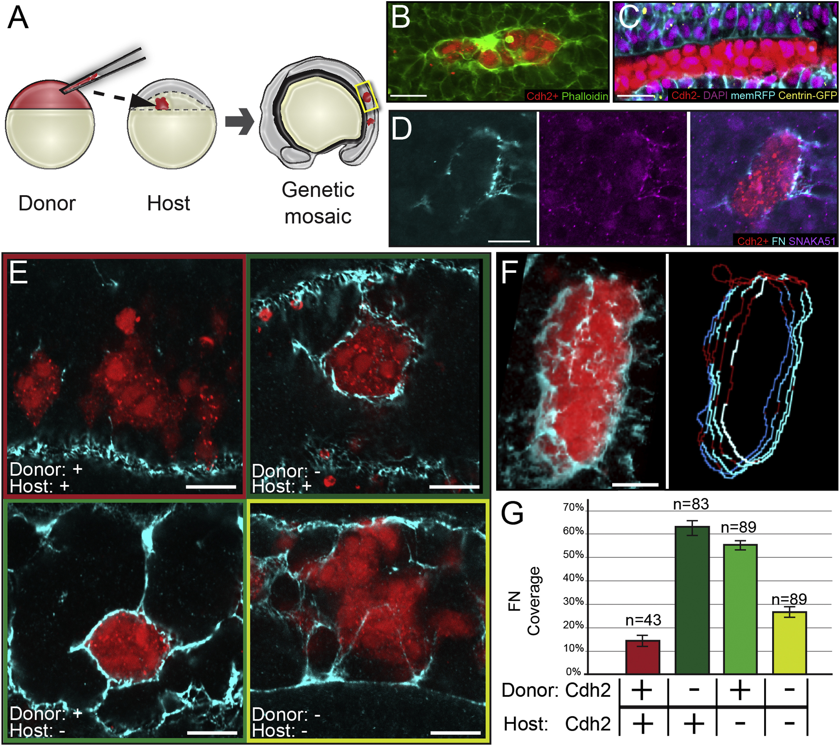
(A) Genetic mosaics were generated by transplanting blastomeres from early blastula-stage donors labeled with fluorescent dextran (rhodamine or Alexa Fluor 647) into later blastula-stage hosts.
(B) Clones were analyzed for F-actin localization via phalloidin staining. 17 of 27 clones exhibited strong cortical actin.
(C) Cdh2-expressing cells polarize toward boundaries with Cdh2-deficient cells. Ten of 12 clones exhibit columnar cell shape, basal nuclei, and apical centrosomes.
(D) Clones were analyzed for Itgα5 activation using the SNAKA51 antibody and FN matrix assembly. Nine of nine clones displayed activated Itgα5 and FN matrix.
(E) Robust FN matrix forms at boundaries between Cdh2-expressing and Cdh2-deficient cells.
(F and G) The percentage of FN coverage of the rhodamine-labeled clones was quantified in multiple z-slices using an ImageJ macro (see Supplemental Experimental Procedures). Pixels positive for FN are blue; pixels negative for FN are red. n, the number of clones examined.
Scale bars, 20 µm. See also Figure S2.