
Fig. 5
|
Fig. 5
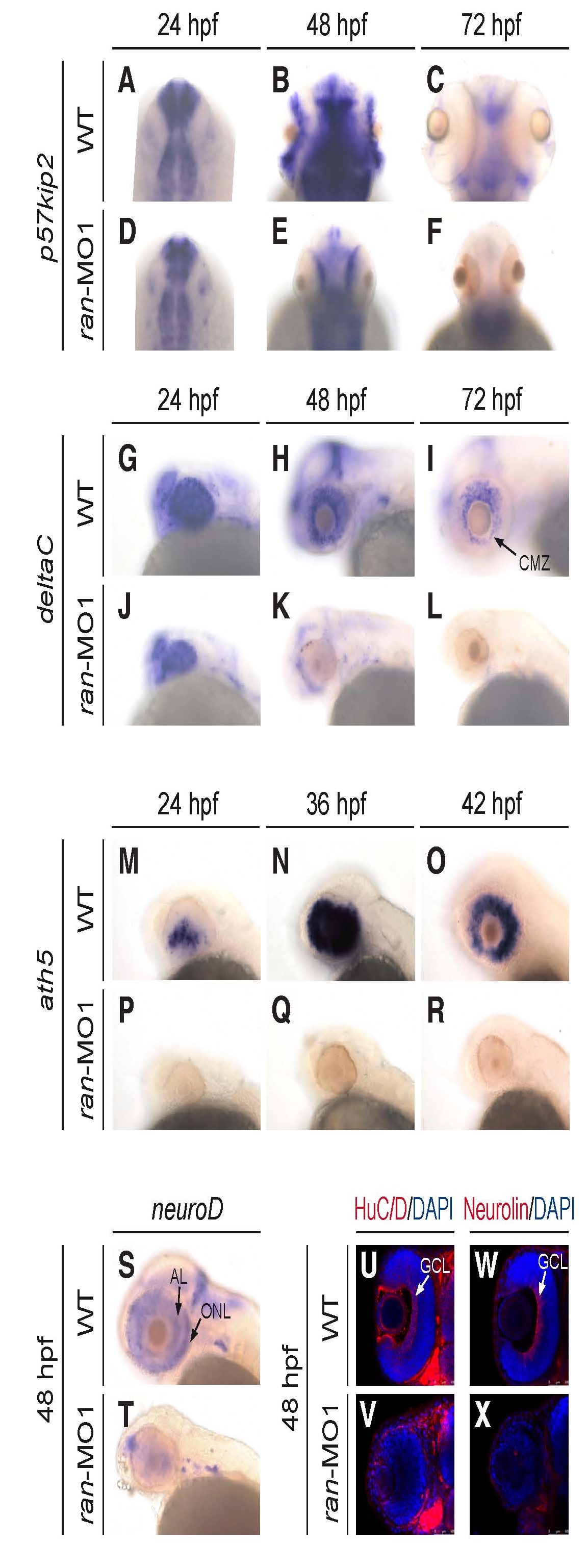
Ran deficiency impaired eye cell differentiation. (A-R) Temporal dynamics of the expression of cell differentiation marker (A-F) p57kip2, (G-L) deltaC and (M-R) ath5 on the eyes of WT and ran-deficient embryos. (S, T) Expression of cell differentiation marker neuroD on the eyes of WT and ran-deficient embryos. (U-X) Expression of the cell differentiation marker (U,V) HuC/D protein and (W,X) Neurolin protein on the eyes of WT and ran-deficient embryos. (A-F) Dorsal view with anterior to the top; (G-T) lateral view with anterior to the left; (U-X) dorsal view with anterior to the top, and only the left eyes are presented to show better detail. AL, amacrine layer; CMZ, ciliary marginal zone; GCL, ganglion cell layer; ONL, outer nuclear layer. All images are representative of at least three repeats with n>25 for each repeat.