
|
Fig. 3
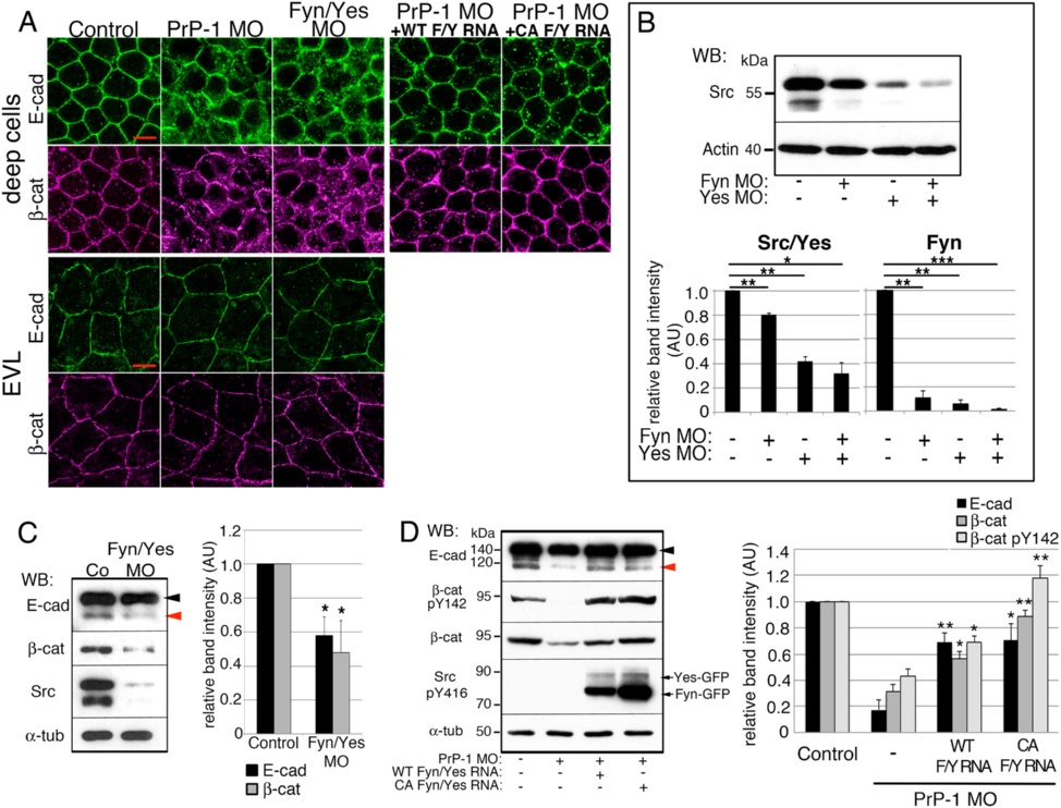
PrP-1 regulates AJs via Fyn and Yes. a. Localization of AJ components (immunofluorescence) in deep (top) and EVL cells (bottom) of 6 hpf PrP-1, Fyn/Yes morphants or PrP-1 morphants co-injected with Fyn/Yes mRNAs. Scale bars = 10 µM. See also Additional file 1: Figure S2. b. Reduction in Src/Yes (65 kDa) and Fyn (50 kDa) total levels in 6 hpf Fyn and Yes single or double knockdowns. c, d. Levels of AJ components in 6 hpf embryo lysates. Activated WT or CA Fyn- and Yes-EGFP were detected with an anti-phospho-Y416 Src antibody. E-cadherin arrowheads as in Fig. 1. WB = Western blot. Densitometric analysis of Western blot bands is expressed in arbitrary units (AU) in b, c and d; average values of three independent experiments ± SEM are shown; statistical significance was assessed using unpaired, two-tailed t-tests; ns = not significant (p > 0.05), * = p ≤ 0.05, ** = p ≤ 0.01, *** = p ≤ 0.001