
|
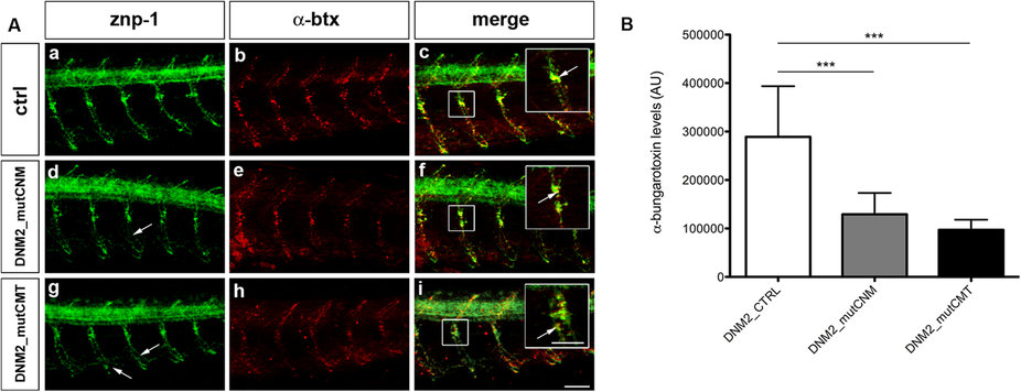
Fig. 5
Primary motor neurons (znp-1) and AChRs (α-btx) in human mRNAs injected embryos.
(A) Co-localization of markers for primary motor neurons (znp-1) and AChRs (α-btx) in 5 spinal hemisegments and somites, in 48 hpf zebrafish embryos. The images are representative of those found in n = 10 embryos for each condition, during 3 distinct experiments. In DNM2_mutCNM-injected embryos (d,e) and DNM2_mutCMT-injected embryos (g,h) primary motor axons migrate normally along the common path (compare a with d and g) with slight pathfinding and shape defects after the choice point (arrows). The merge images suggest that DNM2_mutCNM- and DNM2_mutCMT-injected embryos present fewer α-btx-positive spots than DNM2-controls, with the co-localization signal is reduced in intensity (f,i), compared to control (c), even though znp-1 and α-btx signals co-localize correctly (arrowhead in the enlargement, scale bar = 25 µm). Scale bar = 20 µm. (B) Quantification of α-btx-positive spots in n = 10 complete embryos for each condition. DNM2_mutCNM and DNM2_mutCMT- injected embryos present fewer α-btx-positive spots than controls. Error bars are SEMs. Scale bar = 20 µm.