
Image
|
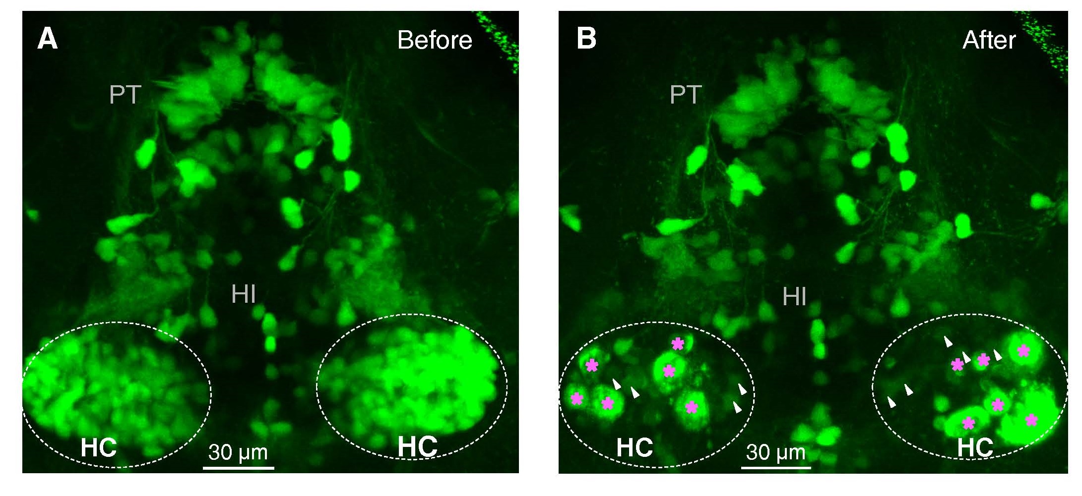
Figure Caption
Fig. S3
Related to Figure 3. Two-Photon Images Taken Before and After Lesion of HC Dopaminergic Neurons. (A and B) Before (A) and after (B) two-photon laser-based lesion of the bilateral HC dopaminergic neurons in a 5-dpf ETvmat2:GFP larva. The pink asterisks indicate bulb-like structures, which suggest successful lesion, and the white arrowheads indicate remaining dopaminergic neurons.
Acknowledgments
This image is the copyrighted work of the attributed author or publisher, and
ZFIN has permission only to display this image to its users.
Additional permissions should be obtained from the applicable author or publisher of the image.
Full text @ Neuron