
|
Fig. 5
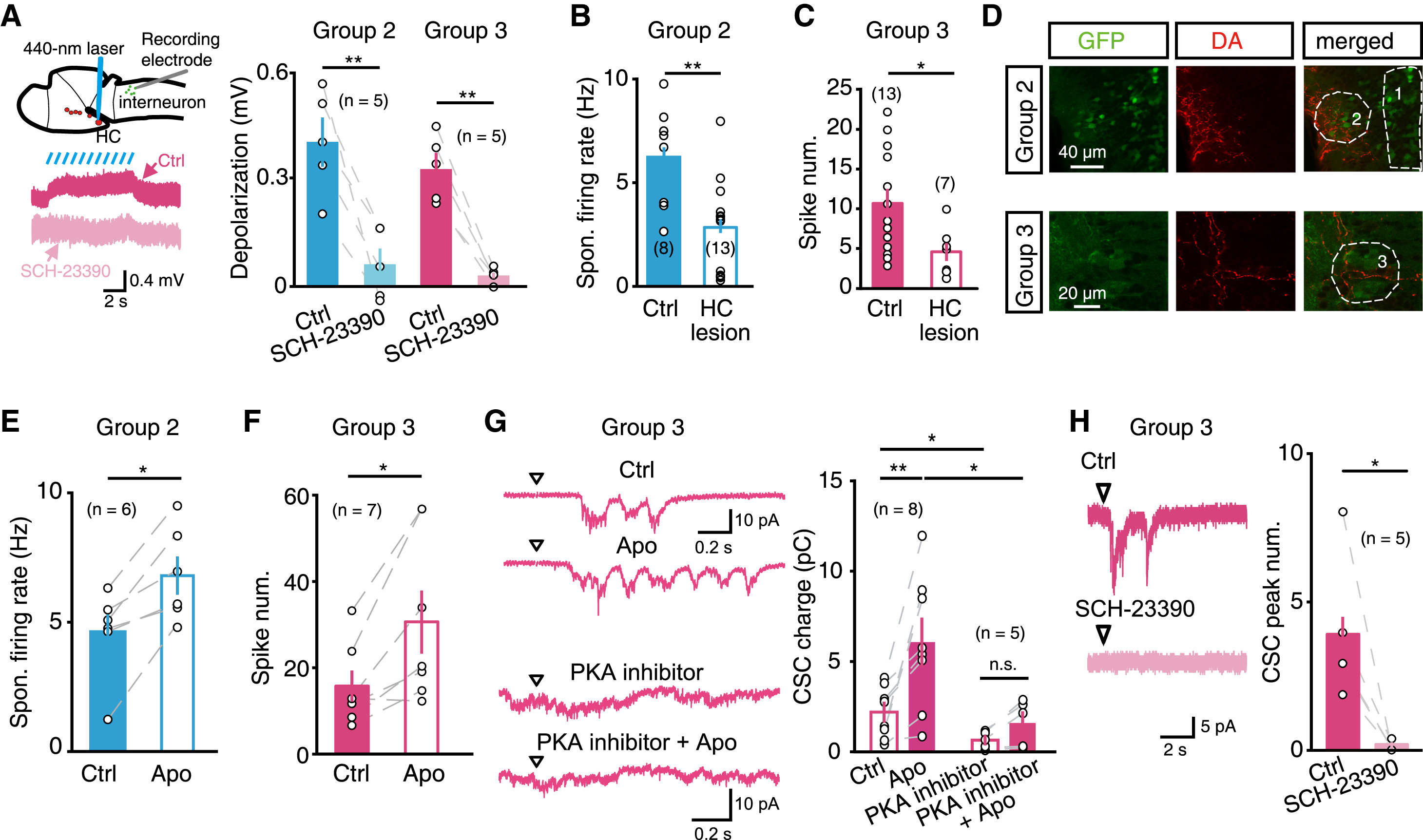
HC Dopaminergic Neurons Directly Modulate Hindbrain Glycinergic Interneurons via D1Rs
(A) Effect of the optogenetic activation of HC dopaminergic neurons on the membrane potential of hindbrain glycinergic interneurons. Left top, schematic of experimental paradigm: in vivo whole-cell recording was performed in glycinergic interneurons, and a 440 nm laser was used to scan the middle layer of the ipsilateral HC in double transgenic Tg (GlyT2:GFP,DAT:ChR2) larvae. Left bottom, sample traces showing that HC optogenetic activation-induced depolarization of glycinergic interneurons was blocked by SCH-23390 application. Right, summary.
(B and C) Effects of HC lesion on the spontaneous firing of the group 2 (B) and flash-evoked firing of the group 3 (C) glycinergic interneurons.
(D) Double immunostaining showing dopamine-immunoreactive fibers in the areas of the groups 2 (top) and 3 (bottom) glycinergic interneurons in a 6 dpf Tg(GlyT2:GFP) larva.
(E and F) Effects of apomorphine (“Apo”) application on the spontaneous firing of the group 2 (E) and flash-evoked firing of the group 3 (F) glycinergic interneurons.
(G) Flash-evoked CSCs in the group 3 glycinergic interneurons before and after application of apomorphine in the absence or presence of intracellular PKA inhibitor. Left, sample traces with the top two obtained from one interneuron and the bottom two from another interneuron. Right, summary.
(H) Effect of local puffing of SCH-23390 on flash-evoked CSCs in the group 3 glycinergic interneurons. Left, sample traces. Right, summary.
*p < 0.05, **p < 0.01; error bars, SEM.
See also Figure S6.