
|
Fig. S11
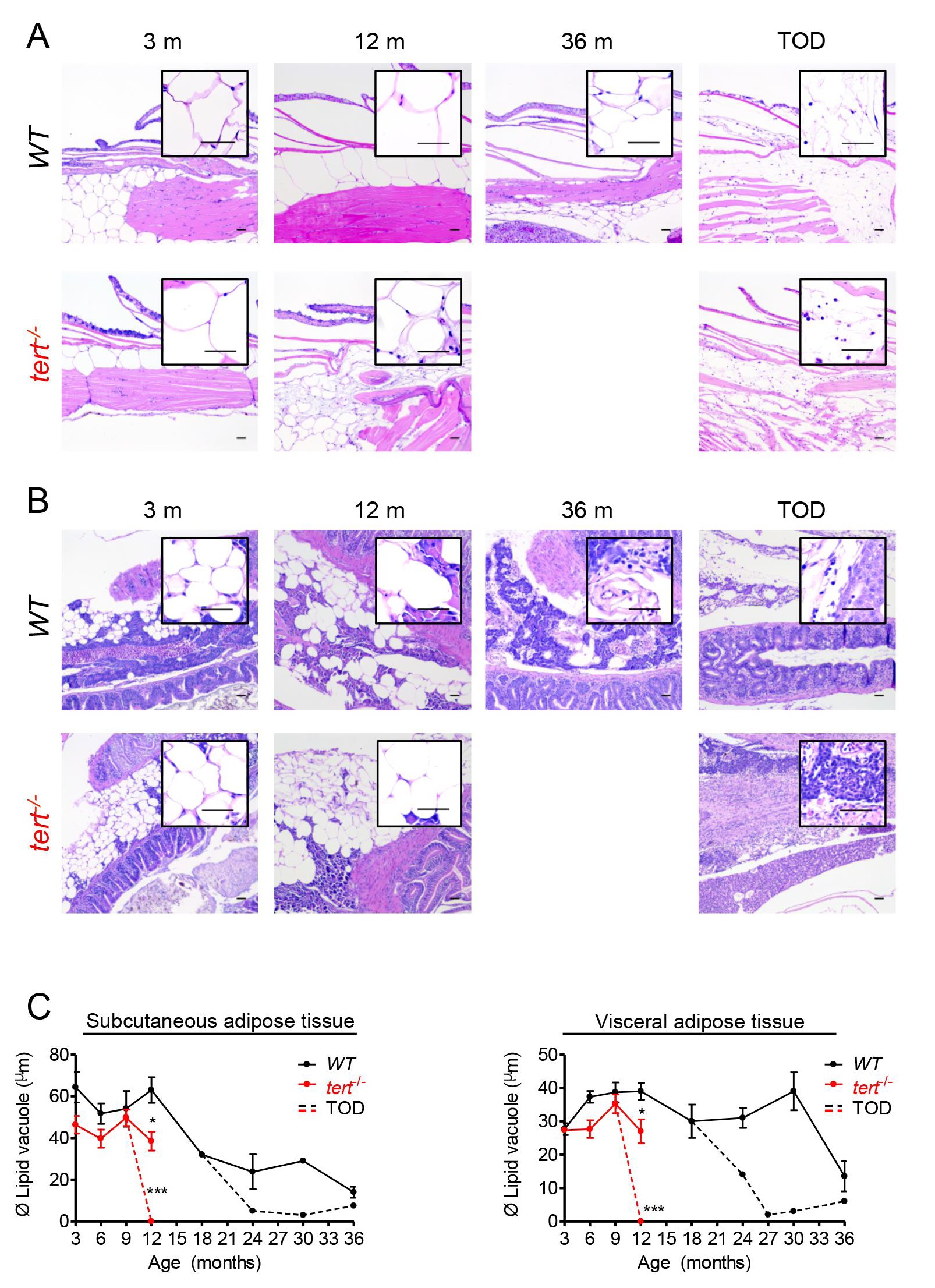
Subcutaneous adipose tissue progressively disappears with aging whereas visceral reserves maintain until later ages.Representative hematoxilin and eosin-stained sections of subcutaneous (A) and visceral (B) adipose tissue depots of WT (3, 12 and 36 months and TOD) and tert-/- siblings (3 and 12 months and TOD), and C) quantification of the adipocyte vacuole diameter at different ages. A) WT zebrafish show a progressive loss of the subcutaneous depot, with age, C) accompanied by a reduction in the adipocytes’ vacuole diameter (adipocytes are ~3.3 times smaller at 36 months vs. 3 months). tert-/- anticipate this phenotype by 12 months of age. With cachexia, both WT and tert-/- zebrafish show complete exhaustion of the subcutaneous adipose tissue depot (at TOD). B) Visceral adipose tissue (peri-pancreatic) is reduced or absent at 36 months in WT zebrafish (when adipocytes are ~3 times smaller than at 3 months) and by 12 months in WT mutants; and similarly to subcutaneous depot, exhaustion of visceral adipose reserves associates with cachexia. N = 3–7 zebrafish per genotype per time point. Time of death (TOD) corresponds to the interval comprising the second and third quartiles of survival (25 to 75%) Scale bar = 50 µm. Data are represented as mean +/- SEM.