
|
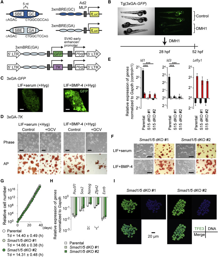
Fig. 3
BMP-SMAD Pathway Is Dispensable for Maintaining Naive Pluripotency
(A) Schematic presentation of BMP reporter constructs. Fragments with GGCGCC (GC) or GGAGCC (GA) sequences together with SBE were triplicated and fused with reporter genes. TK, herpes simplex virus type 1 thymidine kinase gene.
(B) A BMP type I receptor inhibitor, DMH1 (5 µM), attenuated GFP expression in Tg(3xGA-GFP) transgenic fish in vivo. Control, head to the right position; DMH1, head to the left position.
(C) Expression of GFP in 3xGA-GFP cells in LIF+serum or LIF+BMP-4. Scale bar, 40 µm.
(D) Negative selection with ganciclovir (GCV) in 3xGA-TK cells. Upper: Phase contrast. Lower: AP staining. Scale bar, 200 µm.
(E) qRT-PCR analysis to evaluate BMP responsiveness. Cells were stimulated with 50 ng/ml BMP-4 or 50 ng/ml activin A for 1.5 hr. Data represent means ± SEM of three independent experiments. p < 0.001 versus parental mESCs. S15 dKO, Smad1/5 double knockout .
(F) Morphology (left) and AP activity (right) of Smad1/5 dKO lines in LIF+serum and LIF+BMP-4. Scale bar, 200 µm.
(G) Proliferation and doubling time (Td) of parental mESCs and Smad1/5 dKO cell lines maintained in LIF+serum. A representative growth curve data is shown. Td (mean ± SEM) was calculated using three independent experiments.
(H) qRT-PCR analysis of Smad1/5 dKO cell lines cultured in LIF+serum. Data represent means ± SEM of three independent experiments. *p < 0.05, **p < 0.01 versus parental mESCs.
(I) Expression and localization of TFE3 (green), DNA/DRAQ5 (blue), and their merge in Smad1/5 dKO lines in LIF+serum. Scale bar, 20 µm.
See also Figure S2.