
|
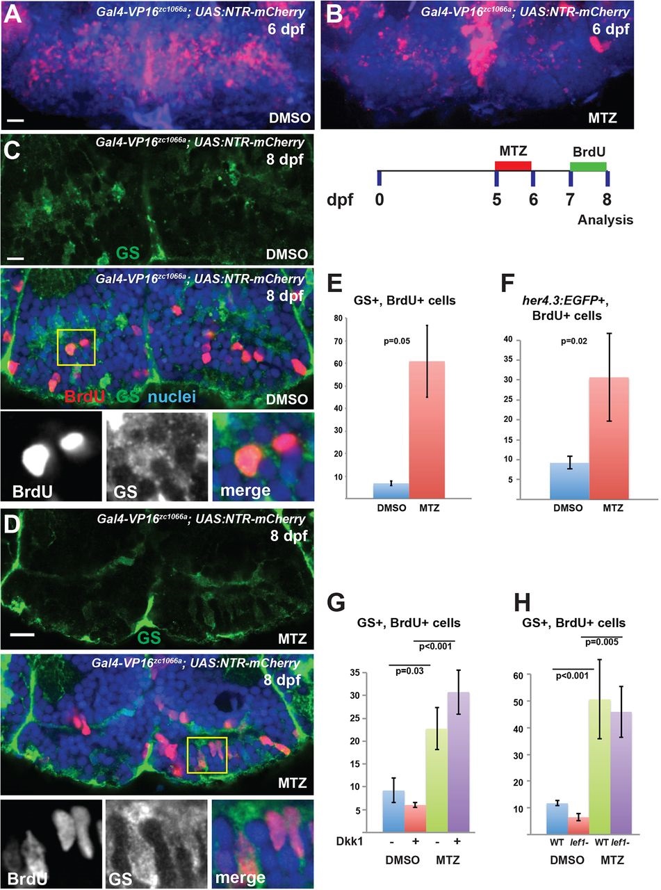
Fig. 4
Hypothalamic radial glia respond to partial ablation by increasing proliferative activity. (A,B) Partial ablation of NTR-mCherry-expressing radial glia (red) by incubation in 1mM MTZ from 5-6dpf; DMSO provides a control. (C-F) After partial ablation from 5-6dpf and BrdU labeling from 7-8dpf (C,D; yellow box indicates region shown beneath), there is a significant increase in the number of BrdU+ radial glia labeled either by GS (C-E) or by her4.3:EGFP (F) expression. (G,H) Inhibition of Wnt signaling by Dkk1 expression (G), or lef1 mutation (H), does not block the increase in BrdU labeling following partial ablation. Images are maximum-intensity z-projections (A,B) or single optical sections (C,D) from ventral views of whole-mount brains. Error bars indicate s.e.m.; n=40 optical sections from four brains for each experiment. Scale bars: 10µm.