
|
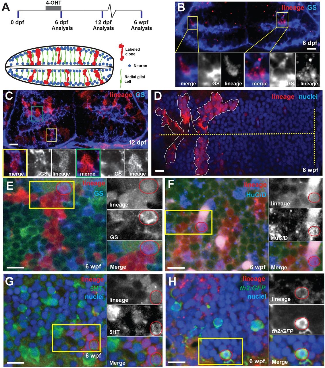
Fig. 2
Hypothalamic radial glia are self-renewing neural progenitors. (A) Timeline and schematic of experiments. Cells expressing 3her4.1:ERT2-Cre-ERT and ubi:loxP-eGFP-loxP-mCherry were genetically labeled by the addition of 5µM 4-OHT from 5-6dpf. Labeled progeny (red) comprise expanding radial units spanning hemispheres of the posterior recess. Processes of GS+ radial glia (green) span the tissue, and nuclei occupy variable positions from the ventricle to the outer edge. Neurons (blue) are located either adjacent to the ventricle or at the outer edge. (B) Immediately following conversion, the few labeled mCherry+ cells are also GS+. Yellow boxes indicate regions shown beneath. (C) Six days after conversion, labeled cells extend radially and include GS+ (yellow box) and GS (green box) cells. (D) Five weeks after 4-OHT addition, discrete groups of mCherry+ cells extend from the ventricle (dashed line). (E-H) mCherry+ progeny 5weeks after recombination include GS+ radial glia (E), HuC/D+ neurons (F), 5HT+ neurons (G) and th2:gfp+ dopaminergic neurons (H). Yellow boxes indicate regions shown beneath, and double-labeled cells are indicated by red circles. Green signal in E,F is ubi:GFP from unconverted cells. Images are single optical sections from ventral views of whole-mount brains. Scale bars: 10µm.