
|
Fig. 4
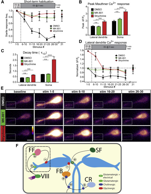
PSP Depression Drives Short-Term Startle Habituation
(A) 500 µM MK-801 (n = 8) and 100 µM strychnine (n = 9) strongly reduce short-term habituation of the startle response compared to DMSO-treated controls (n = 8). A 5-min rest period allows complete recovery to the 31st stimulus.
(B) Peak ΔF/F0 levels in M-cell lateral dendrite and soma are unaffected by MK-801 but are increased by strychnine (p < 0.001, p < 0.0001, t test).
(C) Ca2+ signal decay kinetics are unaltered by MK-801 but are increased by strychnine (p < 0.001, p < 0.0001, t test).
(D) Normalized M-cell lateral dendrite Ca2+ responses are decreased ~35% in DMSO-treated fish during habituation while MK-801 treatment prevented this decrease (p < 0.0001, two-way ANOVA). Responses in strychnine-treated larvae decreased from a higher baseline but remained elevated compared DMSO-treated larvae (p < 0.0001, two-way ANOVA).
(E) Representative images for each block of five stimuli show PSP depression in DMSO compared to MK-801- and strychnine-treated fish. Dashed box highlights lateral dendrite (scale bar, 10 µm).
(F) Diagram of the M-cell circuit including known regulatory inputs. Startle habituation arises from an NMDA- and glycine-receptor dependent mechanism that likely results in enhanced transmission from feedforward (FF) inhibitory neurons to the M-cell and may involve depression of acoustic nerve (VIII) inputs to the M-cell. Inputs from downstream spiral fiber (SF), cranial relay (CR), and feedback inhibitory (FB) neurons are most likely not involved in startle habituation. Excitatory (+) and inhibitory () connections are labeled.
In (A)–(D), error bars indicate SEM.