
|
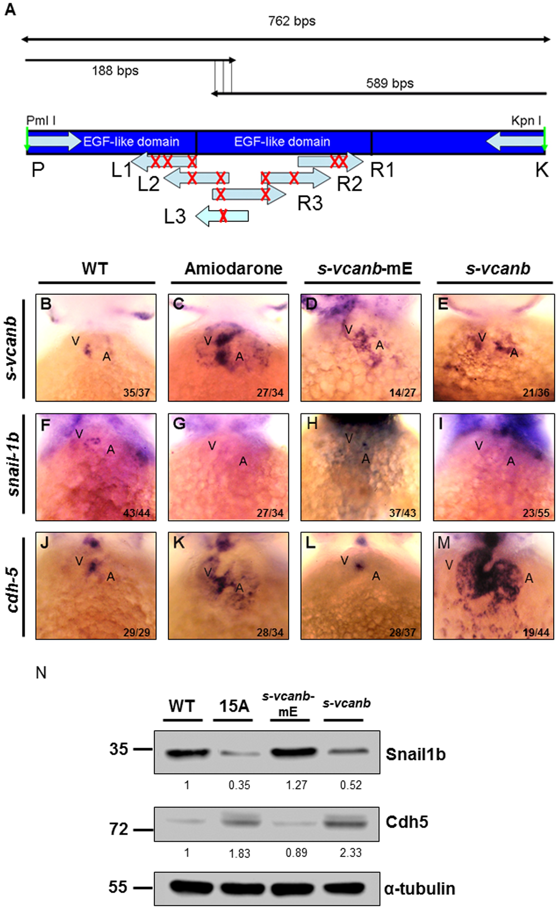
Fig. 3
Amiodarone influences S-vcanb/Snail1b/Cdh5 signaling pathway through the EGF motif of S-vcanb.
(A) Diagram illustrating the mutation sites of S-vcanb at the EGF motif. Six primers (L1–3 and R1–3) containing eight mutation sites (G′C, indicated with X), which abolish the relativity of EGF motif, were used to generate the pS-vcanb-mE. WISH of s-vcanb (B-E), snail1b (F-I) and cdh5 (J-M) in embryos treated with Amiodarone (C, G, K), overexpressed mutation form of s-vcanb-mE (D, H, L), or wild-type s-vcanb (E, I, M). In Amiodarone-treated embryos, s-vcanb (C) and cdh5 (K) were ectopically expressed, but snail1b (G) was lost. In embryos overexpressed in the mutated form of s-vcanb-mE, s-vcanb (D) was increased, but snail1b (H) and cdh5 (L) levels were similar to those of control group (F and J). In embryos overexpressed with the wild-type form of s-vcanb, s-vcanb was increased, but snail1b was lost, while cdh5 was greatly ectopically expressed. (N) Western blot analysis of Snail1b and Cdh5 in wild-type embryos, 72 hpf zebrafish embryos treated with 15 µM Amiodarone from 55 to 72 hpf (15 A), overexpressed mutation form of s-vcanb-mE, and wild-type s-vcanb embryos. Overexpressive wild-type s-vcanb embryos displayed reduced Snail1b and increased Cdh5 patterns. The number of embryos displaying a similar pattern was indicated in each figure. The relative intensities of each protein were as indicated. The α-tubulin was used as an internal control.