
|
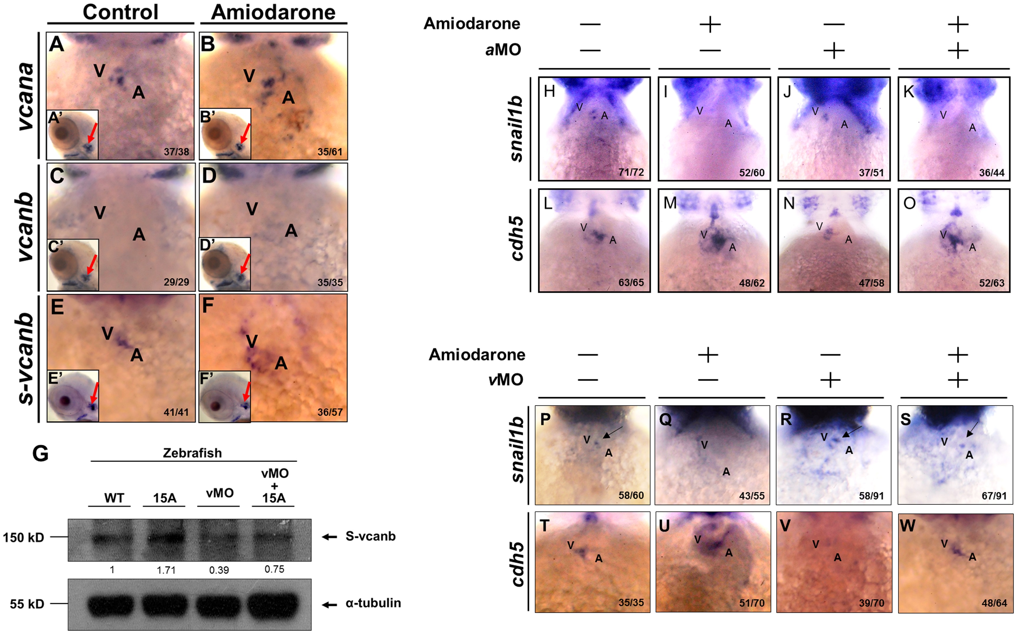
Fig. 2 Reduced Snail1b and increased Cdh5 caused by Amiodarone treatment during valve formation are dependent on S-vcanb.
Using WISH to detect the expression of vcana (A, B), vcanb (C, D) and s-vcanb (E, F) in embryos at 72 hpf was indicated. The head region of embryos treated with either DMSO (Control; A, C, E) or 15 µM Amiodarone (B, D, F) from 55 to 72 hpf. (A′-F′) indicates the head region of each embryo. Red arrows indicate the otic vesicle. The vcana and s-vcanb in the Amiodarone-treated embryos were highlighted. Their ectopic expression occurred only at the heart region. In other tissues, such as otic vesicle, no change was observed. (G) Western blot analysis of S-vcanb protein in wild-type embryos, embryos at 72 hpf after treatment with 15 µM Amiodarone from 55 to 72 hpf (15 A), embryos injected with s-vcanb-MO at one-cell stage (lane 3), and embryos injected with s-vcanb-MO at one-cell stage combined with 15 µM Amiodarone treatment from 55 to 72 hpf (lane 4). The α-tubulin gene served as the internal control. (H-W) Using WISH to detect the expressions of snail1b (H-K, P-S) and cdh5 (L-O, T-W) in WT embryos (H, L, P, T), Amiodarone-treated embryos (I, M, Q, U), vcana-MO-injected embryos (J, N), s-vcanb-MO-injected embryos (R, V), Amiodarone-treatment combined with vcana-MO injection (K, O), and Amiodarone-treatment combined with s-vcanb-MO injection (S, W). V: ventricle, A: atrium. The number of embryos displaying a similar pattern was indicated in each figure.