
|
Fig. S2
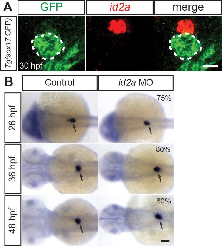
id2a knockdown does not result in a general endoderm-derived organ defect. (A) id2a in situ hybridization (red) combined with anti-GFP immunostaining (green) in Tg(sox17:GFP) embryos reveals that id2a is highly expressed in the interrenal primordium but not in the dorsal pancreas (dashed lines) at 30 hpf. Single confocal section images. (B) id2a MO-injected and control embryos were processed for WISH with the insulin probe, which marks pancreatic beta cells of the dorsal pancreas (arrows). Overall size of the dorsal pancreas appeared unaffected in the MO-injected embryos compared to controls. The percentage of id2a MO-injected embryos exhibiting the representative phenotype shown is indicated in the upper left corner (n=10-20). The remaining percentage of embryos exhibited an intermediate or slightly larger dorsal pancreas phenotype. Arrows point to the dorsal pancreas. Scale bars: 20 µm (A) and 100 µm (B).
Reprinted from Mechanisms of Development, 138 Pt 3, Khaliq, M., Choi, T.Y., So, J., Shin, D., Id2a is required for hepatic outgrowth during liver development in zebrafish, 399-414, Copyright (2015) with permission from Elsevier. Full text @ Mech. Dev.