
IMAGE
Fig. 6
- ID
- ZDB-IMAGE-151229-6
- Publication
- Isaacman-Beck et al., 2015 - The lh3 Glycosyltransferase Directs Target-Selective Peripheral Nerve Regeneration
- All Figures
- Figures for Isaacman-Beck et al., 2015
Image
|
Figure Caption
Fig. 6
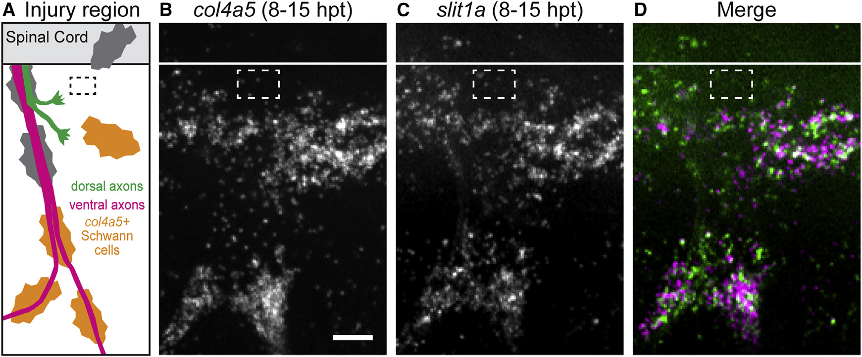
Slit1a Is Upregulated with collagen4a5 after Nerve Transection
(A) Schematic showing approximate region imaged for in situ hybridization (black dashed box, transection site).
(B–D) col4a5 mRNA (B) and slit1a mRNA (C) are co-expressed (D) ventral to the spinal after nerve transection (n = 6 larvae, 27/30 nerves). White line, dorsal aspect of spinal cord; white dashed box, approximate transection site; scale bar, 10 µm.
Acknowledgments
This image is the copyrighted work of the attributed author or publisher, and
ZFIN has permission only to display this image to its users.
Additional permissions should be obtained from the applicable author or publisher of the image.
Full text @ Neuron