
Fig. 1
|
Fig. 1
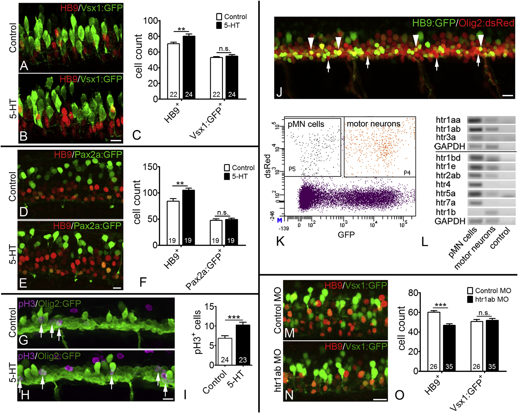
Serotonin Signaling Promotes Embryonic Motor Neuron Generation
Lateral views of spinal cords at 33 hpf are shown.
(A–F) Serotonin (5-HT) treatment (24–33 hpf) increases the number of HB9 immuno-labeled motor neurons but has no influence on vsx1:GFP (A–C) and pax2a:GFP labeled interneurons (D–F) in the same embryos (Student’s t test in C, p = 0.0077; in F, p = 0.002).
(G–I) Serotonin treatment increases the number of dividing (pH3+) olig2:GFP+ pMN progenitor cells (Student’s t test in I; p = 0.0006).
(J–L) Lateral view of a double-transgenic olig2:dsRed/HB9:GFP embryo is shown with red only (arrows, pMN progenitors) and double-labeled (arrowheads, motor neurons) cells indicated in the spinal cord (J). A typical FACS profile is shown (K). In RT-PCR, serotonin receptors show enrichment in pMN progenitor cells, compared to motor neurons (L). GAPDH is used for comparison.
(M–O) Morpholino knockdown of receptor htr1ab reduces the number of HB9+ motor neurons but does not influence the number of vsx1:GFP+ interneurons in the same embryos (Student’s t test; p < 0.0001).
The scale bar in (B) represents 10 µm for (A) and (B), in (E) represents 10 µm for (D) and (E), in (H) represents 15 µm for (G) and (H), and in (N) represents 15 µm for (M) and (N). See also Figure S1.