
|
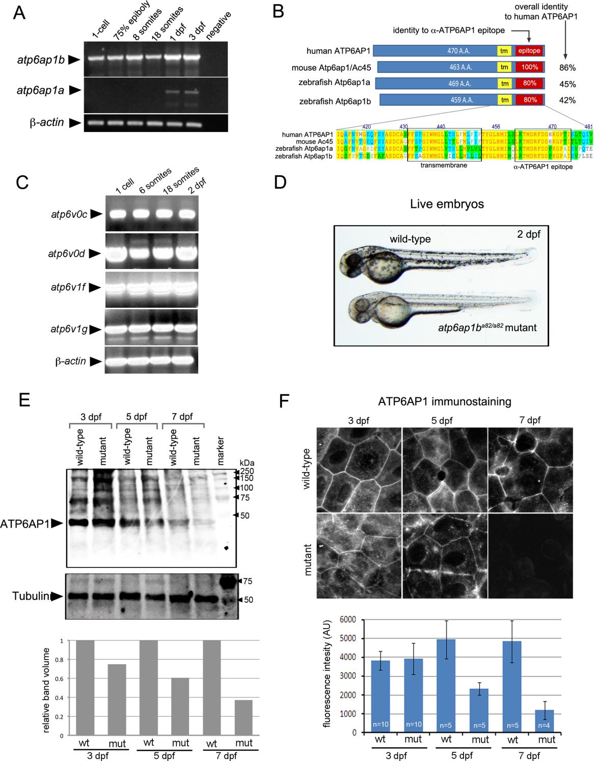
Fig. S1 Expression of the zebrafish V-ATPase accessory protein Atp6ap1b. (A) RT-PCR of temporal expression of atp6ap1b and atp6ap1a through the first three days of development. β-actin was used as a loading control. (B) Schematic diagram and alignment of human, mouse and zebrafish Atp6ap1 proteins. (C) RT-PCR detected maternal mRNA (at the 1-cell stage) of several V-ATPase subunits and then sustained expression through 2dpf. β-actin mRNA was used as a loading control. (D) Homozygous atp6ap1ba82/a82 mutants developed pigmentation phenotypes relative to wild-type siblings. (E) Western blot analysis of Atp6ap1 in wild-type and atp6ap1ba82/a82 mutant embryos at 3, 5 and 7 dpf. A prominent band just under 50 kDa was reduced in mutants. Tubulin was used as a loading control. The graph depicts relative intensities of the Atp6ap1 band from one representative experiment. (F) Fluorescent immunostaining of Atp6ap1 in surface cells of wild-type and atp6ap1ba82/a82 mutant embryos at 3, 5 and 7 dpf. The graph shows the average fluorescent intensity of Atp6ap1 staining.
Reprinted from Developmental Biology, 407(1), Gokey, J.J., Dasgupta, A., Amack, J.D., The V-ATPase accessory protein Atp6ap1b mediates dorsal forerunner cell proliferation and left-right asymmetry in zebrafish, 115-30, Copyright (2015) with permission from Elsevier. Full text @ Dev. Biol.