
|
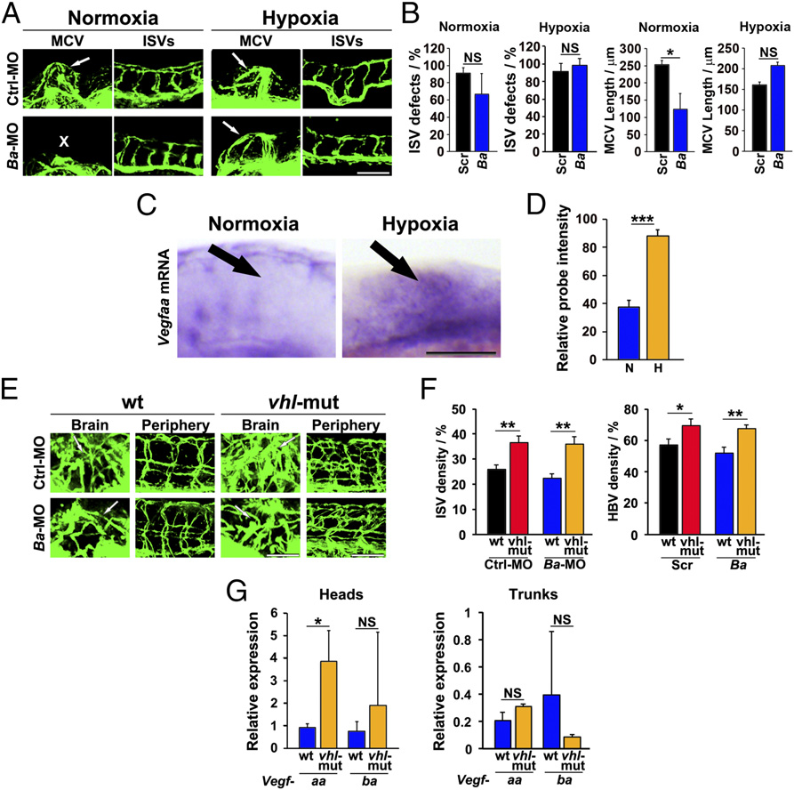
Fig. 7
Hypoxia rescues VEGF-B deficiency-induced vascular defects in zebrafish embryos. (A) Confocal micrographs of 36-hpf stage Fli1a:EGFP zebrafish embryos injected with 0.6 pmol Scrambled (Ctrl) and Vegfba (Ba) morpholinos. The injected embryos were subjected to an E3 medium at normal oxygen tension (normoxia) or to a hypoxic environment where the oxygen tension was reduced to 1.5% (hypoxia). White arrows indicate the MCVs. White X indicates the absence of the MCV. (Scale bar, 100 µm.) (B) Quantification of ISV defects and MCV lengths at the 36-hpf stage of Fli1a:EGFP zebrafish embryos injected with 0.6 pmol Scrambled (Ctrl) or Vegfba (Ba) morpholinos. The injected embryos were subjected to an E3 medium under normoxia or hypoxia conditions (n = 11–21 embryos/group). NS, nonsignificant. *P < 0.05. (C) Detection of Vegfaa mRNA by in situ hybridization of 36-hpf stage embryos exposed to an E3 medium under normoxia or hypoxia. Black arrows indicate the MCV area. (Scale bar, 100 µm.) (D) Quantification of in situ hybridization positive signals under normoxia (N) and hypoxia (H) conditions (n = 35 embryos/group). ***P < 0.001. (E) Confocal micrographs of Fli1a:EGFP-wt (wt) or Fli1a:EGFP-vhl-mutant (vhl-mut) 120-hpf zebrafish embryos injected with 0.6 pmol scrambled (Ctrl) or Vegfba (Ba) morpholinos. White arrows indicate the brain vasculatures. (Scale bar in , 50 µm; bar in periphery, 100 µm.) (F) Quantification of ISV and brain vascular (HBV) density in Fli1a:EGFP-wt (wt) or Fli1a:EGFP-vhl-mutant (vhl-mut) 120-hpf zebrafish embryos injected with 0.6 pmol scrambled (Ctrl) or Vegfba (Ba) morpholinos (n = 9–17 embryos/group). *P < 0.05; **P < 0.01. (G) qPCR analysis of the relative expression of Vegfaa or Vegfba mRNA levels in heads and trunks of 120-hpf Fli1a:EGFP-wt (wt) or Fli1a:EGFP-vhl-mutant (vhl-mut) zebrafish embryos (60–150 embryos/group). NS, nonsignificant. *P < 0.05.