
Image
|
Figure Caption
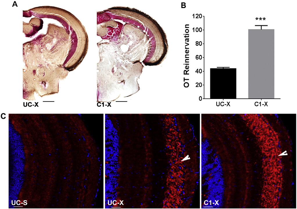
Fig. 5
Blocking TRβ receptor accelerates OT reinnervation. A. Brown staining represents biocytin labeled RGC axons. B. Quantification of the area covered by RGC axons in the OT of UC and C1 fish. Data represent mean ± SEM (n = 6 animals/group). ***p < 0.0001 by unpaired t-test. C. Immunofluorescent detection of Gap43 protein (red) in RGC axons (arrow) arriving in the OT. Cell nuclei are stained with DAPI (blue). Pictures are representative images for n = 4 animals/group. Scale bar - 20 µm. For all panels: UC-untreated control, C1-compound 1 treated, S-sham operated, X-optic nerve crushed, OT-optic tectum.
Acknowledgments
This image is the copyrighted work of the attributed author or publisher, and
ZFIN has permission only to display this image to its users.
Additional permissions should be obtained from the applicable author or publisher of the image.
Reprinted from Molecular and cellular neurosciences, 68, Bhumika, S., Lemmens, K., Vancamp, P., Moons, L., Darras, V.M., Decreased thyroid hormone signaling accelerates the reinnervation of the optic tectum following optic nerve crush in adult zebrafish, 92-102, Copyright (2015) with permission from Elsevier. Full text @ Mol. Cell Neurosci.