
|
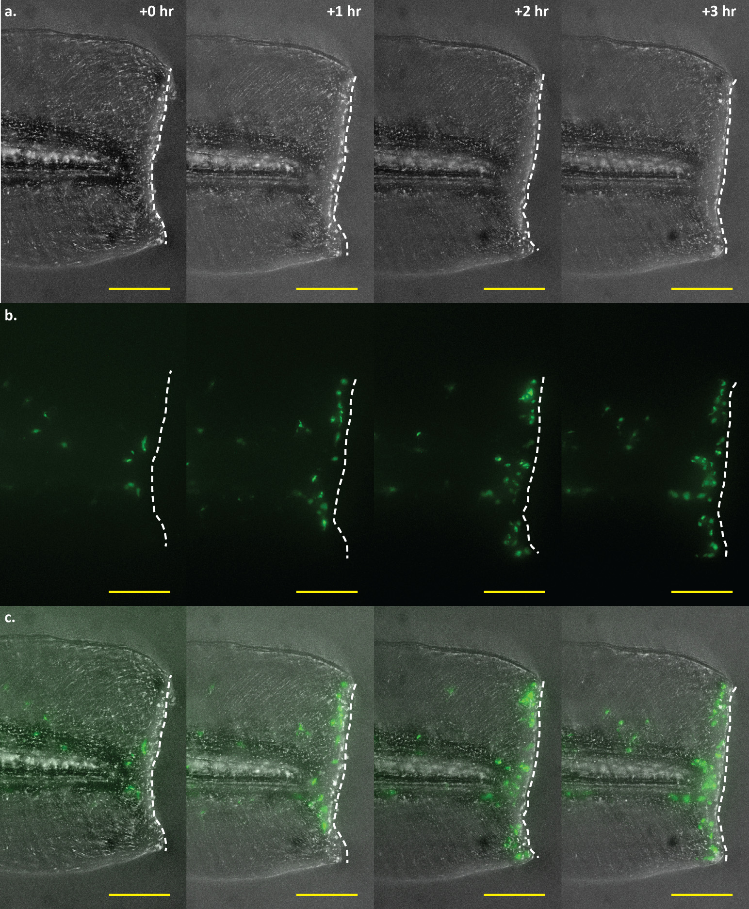
Fig. 3
Time-lapse imaging of a tail wound of an mfap4:dLanYFP-CAAX transgenic larva.
Time-lapse imaging of macrophage recruitment to the site of a wound at the posterior end of the larval tail. Macrophage movements may be visualized in real-time via fluorescence imaging of mfap4 transgenic larvae. Shown here are four representative frames of such an experiment, showing macrophage recruitment to the wound edge at 0, 1, 2, and 3 hours post-wounding. a. Brightfield detailing the extent and location of the wound. The wound edge is highlighted as a dashed white line. b. Macrophages visualized via dLanYFP fluorescence. The number of cells present proximal to and along the site of the wound can be seen increasing over time as macrophages track toward the damaged tissue. c. Merge of brightfield and fluorescence channels. Scale bars = 100 µm. Images representative of 12 animals from two independent biological replicates.