
Image
|
Figure Caption
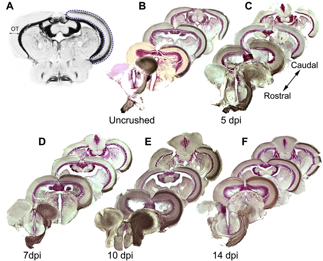
Fig. 1
A. Selection of the white matter area of the optic tectum (OT, blue dotted line) for quantification of the biocytin signal. B–F. Innnervation of the OT by RGC axons in uninjured fish and reinnervation of the OT by regenerating RGC axons at 5, 7, 10 and 14 dpi. Brain sections are arranged from rostral to caudal, the side innervated by the crushed ON on the right. By 5 dpi biocytin labeled regenerating RGC axons (brown) started to enter the OT. At 7 dpi they arrived approximately halfway through the OT. At 10 and 14 dpi, the biocytin labeling matches that observed in uncrushed fish.
Acknowledgments
This image is the copyrighted work of the attributed author or publisher, and
ZFIN has permission only to display this image to its users.
Additional permissions should be obtained from the applicable author or publisher of the image.
Reprinted from Molecular and cellular neurosciences, 68, Bhumika, S., Lemmens, K., Vancamp, P., Moons, L., Darras, V.M., Decreased thyroid hormone signaling accelerates the reinnervation of the optic tectum following optic nerve crush in adult zebrafish, 92-102, Copyright (2015) with permission from Elsevier. Full text @ Mol. Cell Neurosci.