
IMAGE
Fig. S1
- ID
- ZDB-IMAGE-151113-6
- Publication
- Xu et al., 2015 - Temporal-Spatial Resolution Fate Mapping Reveals Distinct Origins for Embryonic and Adult Microglia in Zebrafish
- All Figures
- Figures for Xu et al., 2015
Image
|
Figure Caption
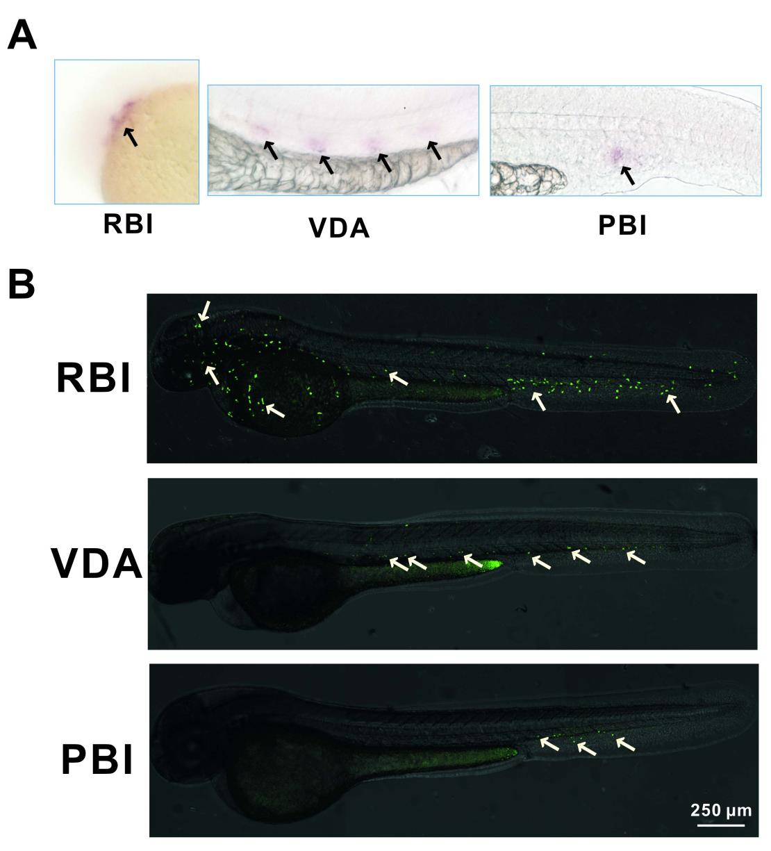
Fig. S1
The IR-LEGO-CreER-loxP System Induces Specific GFP Expression in the Embryos Treated with Infrared Irradiation and 4-OHT.
(A) High magnification of a less exposed staining of CreER in situ hybridization in RBI, VDA and PBI regions.
(B) Digitally reconstituted confocal images reveal GFP signals (indicated by arrows) in the embryos one day after heat shock in the RBI, VDA and PBI regions and 4-OHT treatment.
Acknowledgments
This image is the copyrighted work of the attributed author or publisher, and
ZFIN has permission only to display this image to its users.
Additional permissions should be obtained from the applicable author or publisher of the image.
Reprinted from Developmental Cell, 34, Xu, J., Zhu, L., He, S., Wu, Y., Jin, W., Yu, T., Qu, J.Y., Wen, Z., Temporal-Spatial Resolution Fate Mapping Reveals Distinct Origins for Embryonic and Adult Microglia in Zebrafish, 632-641, Copyright (2015) with permission from Elsevier. Full text @ Dev. Cell