
|
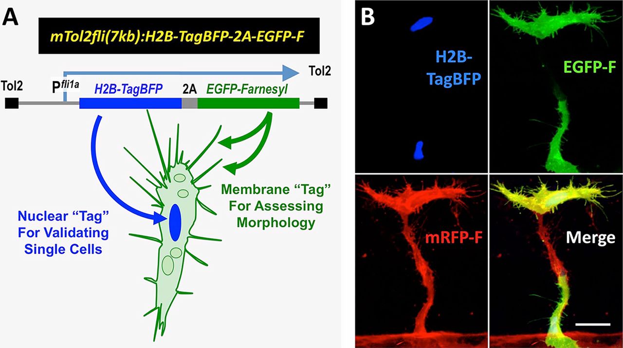
Fig. 2
Design and validation of an expression cassette for analyzing the morphology and dynamics of individual ECs within developing vessels. (A) The Tol2(fli1a:H2B-TagBFP-p2A-egfp-F) transgene construct for simultaneously marking endothelial nuclei (with H2B-TagBFP) and EC internal and plasma membranes (with eGFP-farnesyl). (B) Confocal micrograph of a growing trunk ISV/DLAV segment in a 32hpf Tg(kdrl:mRFP-F)y286 embryo injected with a Tol2(fli1a:H2B-TagBFP-p2A-egfp-F) transgene, showing blue, green and red fluorescent channels and all three merged. This segment contains two individual transgene-labeled ECs (as indicated by the presence of a single H2B-TagBFP nucleus in each). eGFP-F fluorescence is concentrated on the cell surface as well as on internal membranes, although not readily evident in this low-magnification reconstructed image. Scale bar: 20µm.