
|
Fig. 6
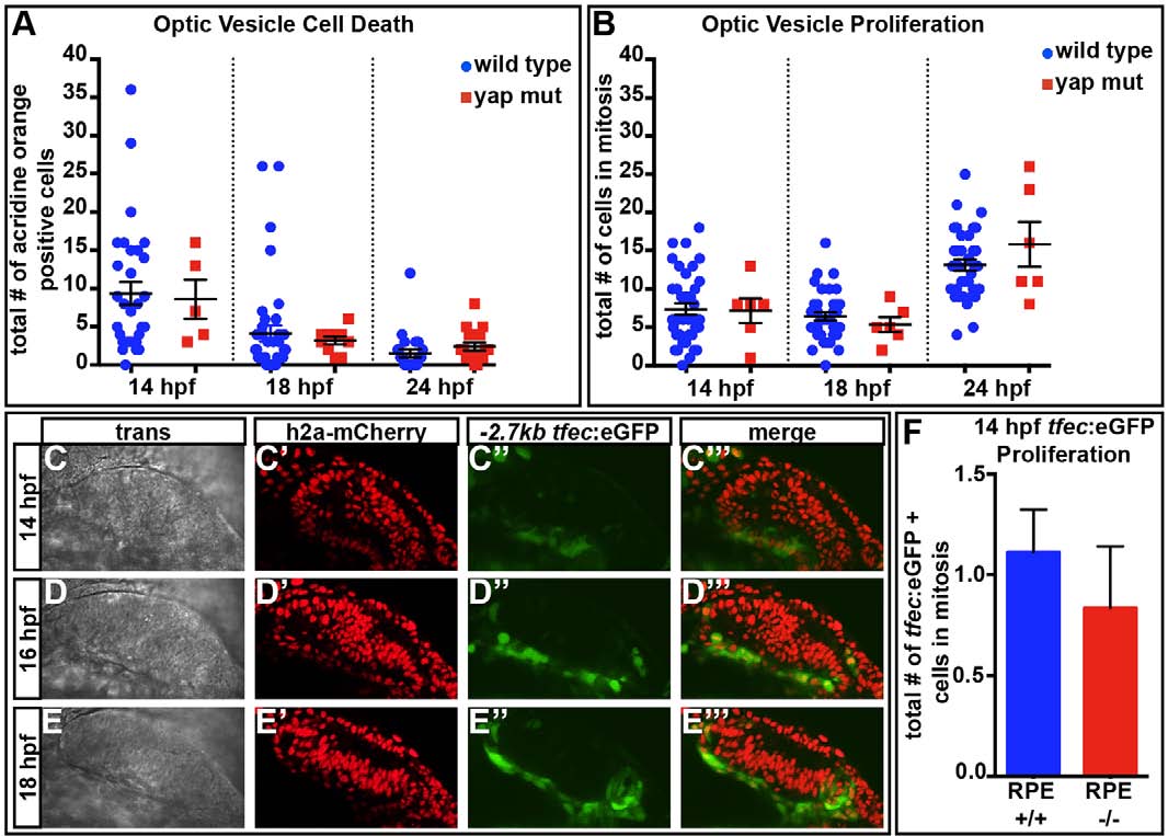
Cell death and proliferation are normal in yap-/- eyes. (A) The numbers of dying cells identified by Acridine Orange staining do not differ in yap-/- mutants at 14 (P=0.8465), 18 (P=0.6542) or 24 (P=0.2558) hpf as compared with wild type. Numbers of eyes analyzed: 14hpf, wt n=31, yap-/- n=5; 18hpf, wt n=39, yap-/- n=10; 24hpf, wt n=24, yap-/- n=16. (B) Eye field mitotic cell counts do not differ between yap+/? (wt) and yap-/- at 14 (P=0.9205), 18 (P=0.4329) and 24 (P=0.2222) hpf. Numbers of eyes analyzed: 14hpf, wt n=38, yap-/- n=6; 18hpf, wt n=38, yap-/- n=6; 24hpf, wt n=36, yap-/- n=6. (C-E′′′) Time-course of expression of tfec:eGFP in prospective RPE cells. (F) Mitotic cell counts of tfec:eGFP+ cells do not differ between wild-type and yap-/- eyes at 14hpf (P=0.5408). Numbers of eyes analyzed: wt n=18, yap-/- n=6. P-values were obtained using an unpaired t-test with equal s.d. Error bars indicate s.e.m. Wild type included full RPE coverage, whereas yap-/- showed some RPE loss.