
|
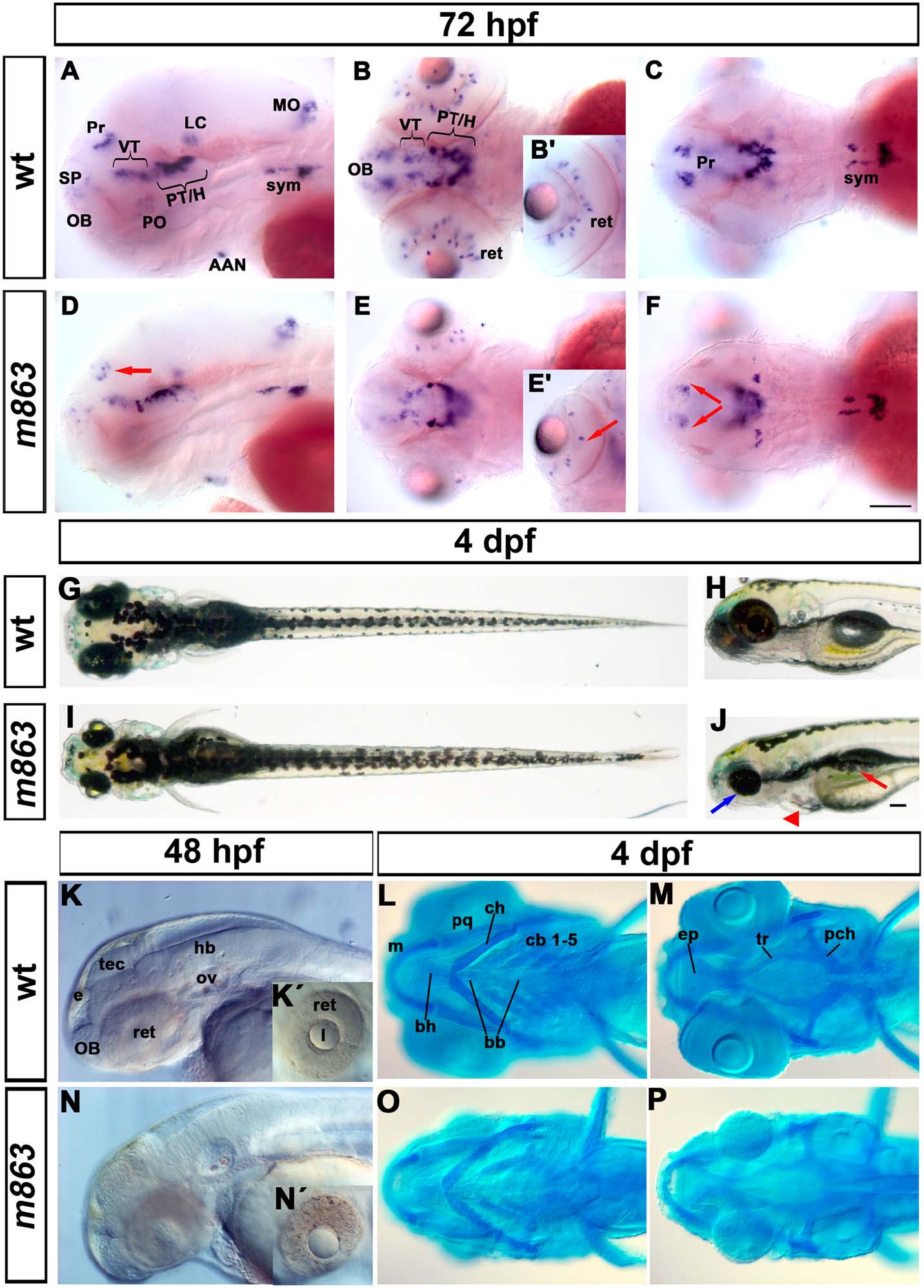
Fig. 1
Phenotype of m863 mutant embryos.
(A-F) Reduction of th-expressing dopaminergic neurons in the pretectum and retina of m863 mutants at 72hpf. (A-C) Lateral (A) and dorsal (B-C) views of th expression pattern in wild type siblings. (D-F) Lateral (D) and dorsal (E-F) views of th expression pattern in homozygous mutant m863 larvae. Arrows indicate affected DA groups in the pretectum (D, F) and retina (E′). (G-J) Morphological phenotype of live m863 mutants at 4 dpf. Dorsal (G) and lateral (H) views of wild type larvae. Dorsal (I) and lateral (J) views of homozygous m863 mutants. Compared to wild type siblings, m863 mutants displayed smaller eyes (blue arrow), flattened and smaller head, edema (red triangle), and defective swim bladder (red arrow), but normal body length. (K, N) Lateral views of live wild type (K) and homozygous m863 mutants (N) at 48 hpf. Granular tissue appearance in the midbrain region and retina of mutants indicated elevated cell death. (L,M,O,P) Ventral views of Alcian blue staining of head cartilage in wild type (L, M) and homozygous m863 mutants (O, P) at 4 dpf. The cartilaginous head skeleton of mutants was smaller and underdeveloped compared to wild type siblings. Abbreviations used: Catecholaminergic groups: AAN, arch-associated neurons (noradrenergic); H, hypothalamus; LC, locus coeruleus; MO, medulla oblongata (noradrenergic); OB, olfactory bulb; PO, preoptic region; Pr, pretectum; PT, posterior tuberculum; SP, subpallium; sym, sympathetic neurons (catecholaminergic); VT, ventral thalamus. Cartilage structures: bb, basibranchial; bh, basihyal; cb, ceratobranchials; ch, ceratohyal; ep, ethmoid plate; m, Meckel’s cartilage; pch, parachordal; pq, palatoquadrate; tr, trabecula. Anterior towards the left. Scale bar: 100 µm