
Fig. 6
|
Fig. 6
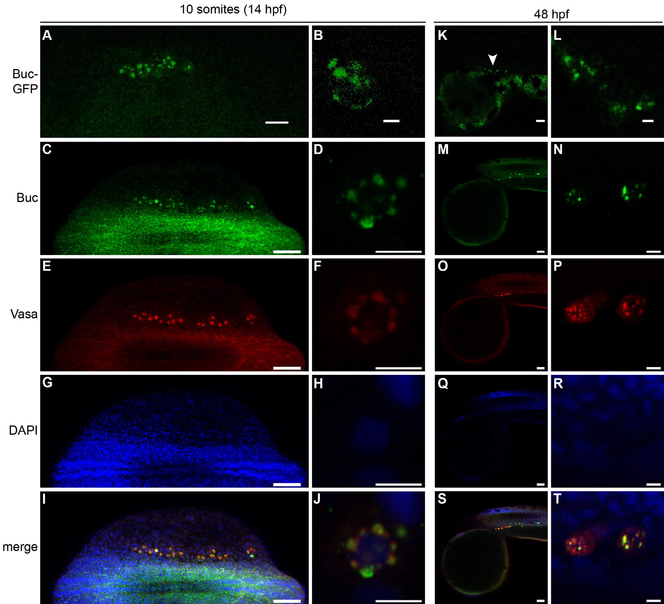
Buc is expressed in larval stages. (A–J) Confocal images of 10-somite stage embryos (14 hpf) in dorsal view, anterior to the left. Only the right half was magnified for imaging (A, C, E, G, I). Magnification of single PGCs at 10-somite stage (B, D, F, H, J). (A, B) Living embryos showing Buc-GFP in germ cells (A), forming perinuclear granules (B). (C–J) Wild-type embryos labeled with Buc-antibody (C, D; green) in germ cells (E, F; Vasa in red) and nuclei (G, H; labeled blue with DAPI). The merge (I, J) shows the colocalization of Buc in Vasa-positive, perinuclear granules. (K–T) Confocal images of 48 hpf embryos in lateral view, anterior to the left. Buc-GFP is expressed in living germ cells (K, L; white arrowhead). (M–T) Buc antibody labeling confirms the co-expression of endogenous Buc (M, N; green) in wild-type embryos with the PGC marker Vasa (O–T; red). Scale bar: 5 µm (B, D, F, H, J, L, N, P, R, T); 50 µm (A, C, E, G, I, K, M, O, Q, S).
Reprinted from Gene expression patterns : GEP, 18(1-2), Riemer, S., Bontems, F., Krishnakumar, P., Gömann, J., Dosch, R., A functional Bucky ball-GFP transgene visualizes germ plasm in living zebrafish, 44-52, Copyright (2015) with permission from Elsevier. Full text @ Gene Expr. Patterns