
Image
|
Figure Caption
Fig. 1
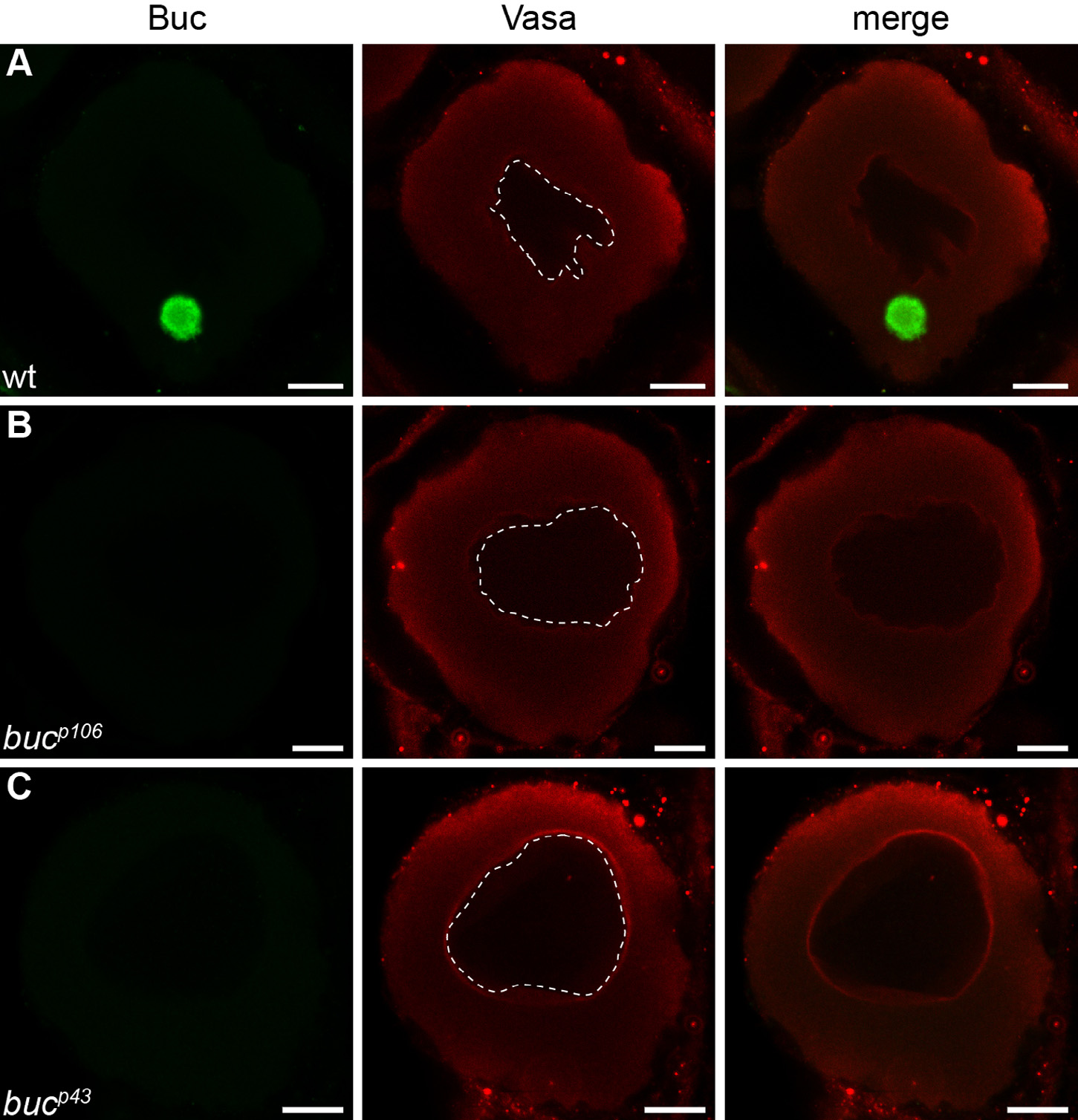
Buc antibody specificity. Confocal images of stage IB oocytes. Buc (green) labels the Balbiani body in wild type oocytes (A) but not in mutants bucp106 (B) and bucp43 (C). By contrast, perinuclear Vasa (red) is not changed. Dashed line outlines the nucleus. Lateral views, animal to the top. Scale bar: 10 µm.
Figure Data
Acknowledgments
This image is the copyrighted work of the attributed author or publisher, and
ZFIN has permission only to display this image to its users.
Additional permissions should be obtained from the applicable author or publisher of the image.
Reprinted from Gene expression patterns : GEP, 18(1-2), Riemer, S., Bontems, F., Krishnakumar, P., Gömann, J., Dosch, R., A functional Bucky ball-GFP transgene visualizes germ plasm in living zebrafish, 44-52, Copyright (2015) with permission from Elsevier. Full text @ Gene Expr. Patterns