
|
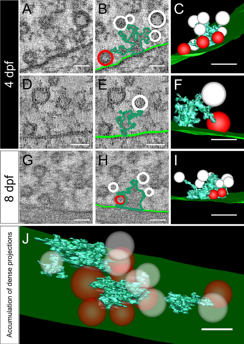
Fig. 6
Dense projections link synaptic vesicles to the presynaptic membrane. Shown are tomograms of dense projections in 4-dpf (A,D) and 8-dpf (G) zebrafish. They were segmented (B,E,H) and rendered into 3D models (C,F,I). Light green, presynaptic membrane; dark green, dense projection; white, synaptic vesicles; red, docked vesicles. All segmentations and 3D models show only the vesicles directly connected to dense projections. A-C and D-F: Typical dense projections in 4-dpf larvae. G-I: Dense projections found in 8-dpf larvae. J: Same tomogram as in D but showing all dense projections found. For a better overview, all vesicles are shown as transparent (J). A magenta/green version of this figure is available as Supporting Information Figure 2. Scale bars = 50 nm in in A,B,D-H,J; 100 nm in C,I.