
|
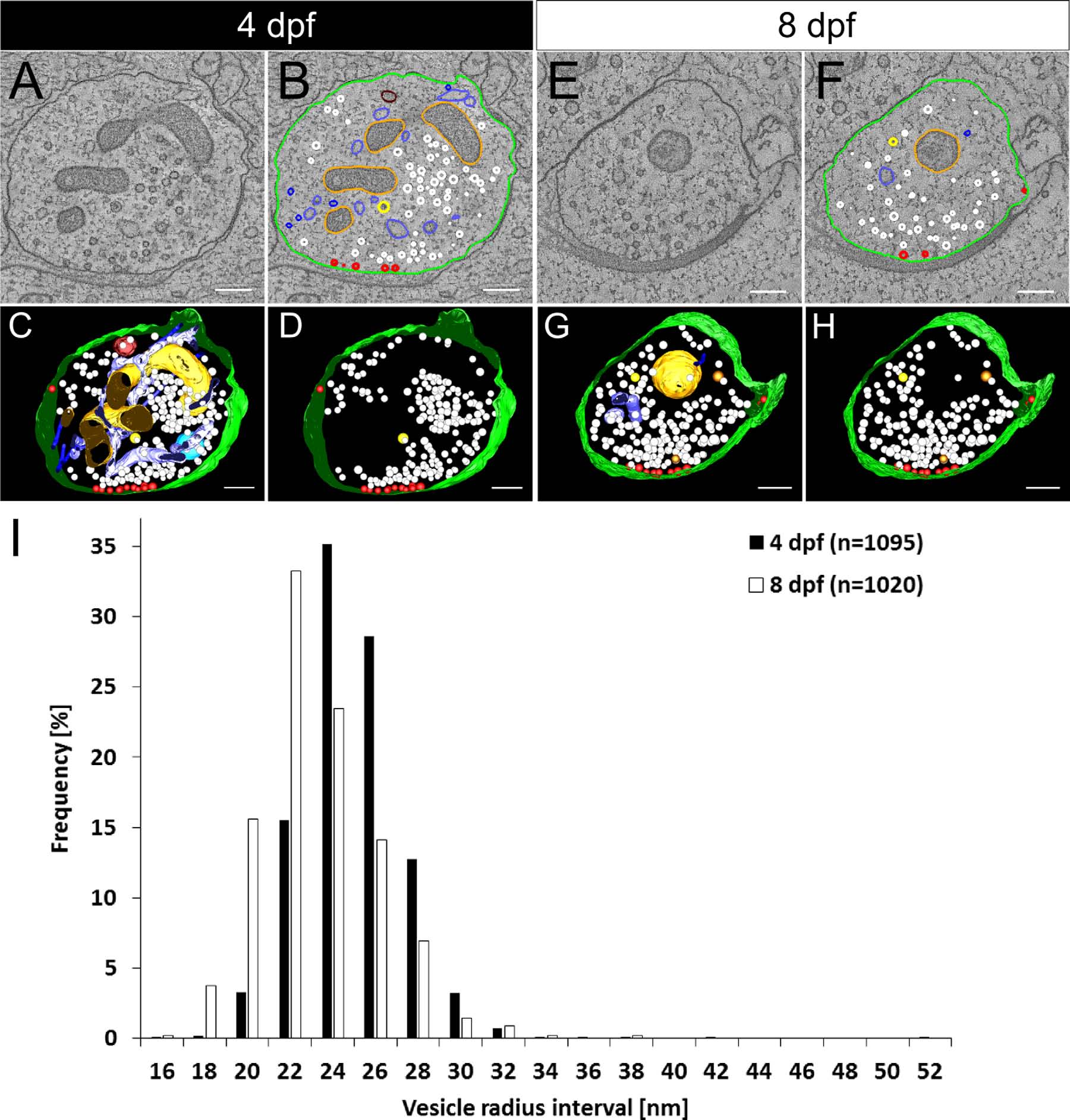
Fig. 3
Differences in size of the synaptic vesicles in NMJs from 4-dpf and 8-dpf zebrafish larvae. Tomograms of NMJs from 4-dpf (A) and 8-dpf (E) zebrafish larvae were taken and the structures were annotated (B,F). In the synapse, synaptic vesicles (white), docked vesicles (red) at the cell membrane (light green), mitochondria (orange), endoplasmic reticulum (light blue), endosomal structures (dark brown), microtubules (dark blue), two types of dense-core vesicles (brown and yellow), and large multivesicular bodies (turquois) were found. 3D models are shown either with all structures (C,G) or with only the vesicle pools of the synapses visible (D,H). The measurements of the sizes of the synaptic vesicles pools at 4 and 8 dpf (I) shows that in 4-dpf zebrafish larvae the radius of synaptic vesicles (23.9 ± 2.5 nm, n = 1,095 vesicles, mean ± SD) is highly significantly larger than that in 8-dpf zebrafish larvae (22.2 ± 2.7 nm, n = 1,020 vesicles, mean ± SD). Scale bars = 200 nm.