
|
Fig. 4
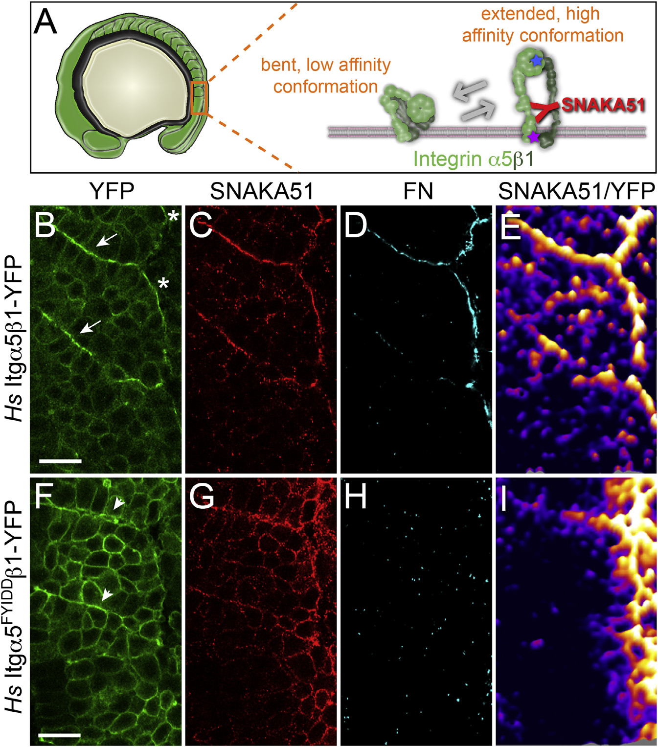
Itgα5 Adopts the Active, Extended Conformation Independently of Ligand Binding
(A) A schematic of the bent and extended Itgα5β1. The SNAKA51 antibody recognizes an epitope on the extracellular domain of Itgα5 that is exposed when the Itgα5β1 adopts the extended, active conformation (see also Figure S2). The blue star represents the five amino acid substitutions in Itgα5FYIDD that eliminate ligand binding. The magenta star represents the two amino acid substitutions in Itgα5GAAKR that promote adoption of the extending active conformation.
(B–E) Confocal images of MZ itgα5-/- mutant embryos expressing Hs Itgα5β1-YFP (B) immunostained with the conformation-specific anti-Hs Itgα5 antibody SNAKA51 (C) and anti-FN antisera (D). The arrows in (B) indicate somite borders and the asterisks denote the lateral surface of the tissue. There were six experiments that were performed with n = 55 embryos analyzed. (E) To normalize the SNAKA51 signal to the level of YFP, a ratiometric topological heat map was generated. The warmer colors indicate higher relative levels of extended, active Itgα5β1. The same pattern of Itg±5 activation is observed in the absence of BiFC when using Hs Itgα5-GFP for ratio imaging (data not shown).
(F–I) Shows confocal images of a parallel analysis of MZ itgα5-/- mutant embryos expressing Hs Itgα5FYIDD ligand binding deficient Integrin. The arrowheads in (F) indicate ephemeral Itgα5FYIDDβ1 clustering. There were six experiments that were performed with n = 52 embryos analyzed.
In (B)–(I), anterior is up and lateral is right. The scale bars represent 20 µm.
Reprinted from Developmental Cell, 34(1), Jülich, D., Cobb, G., Melo, A.M., McMillen, P., Lawton, A.K., Mochrie, S.G., Rhoades, E., Holley, S.A., Cross-Scale Integrin Regulation Organizes ECM and Tissue Topology, 33-44, Copyright (2015) with permission from Elsevier. Full text @ Dev. Cell