
|
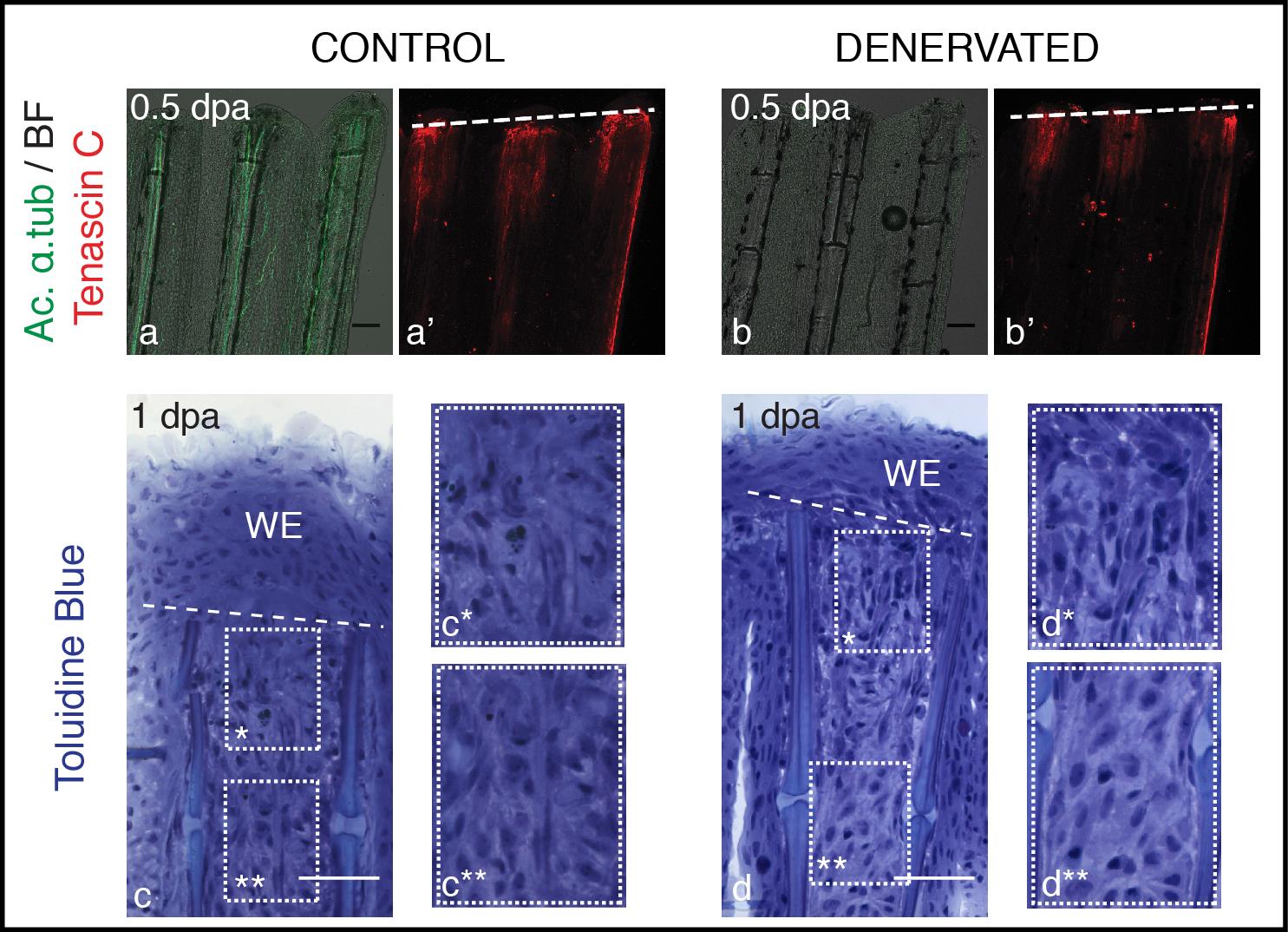
Fig. S2
Mesenchymal tissue disorganization after amputation of denervated fins. a,b) Staining for Tenascin C and ac. α-tub in whole mount fins at 0.5 dpa. Tenascin C is present in the mesenchymal tissue under the amputation plane, both in control and denervated fins. c,d) Toluidine Blue histology in longitudinal sections. Longitudinal sections stained with toluidine blue show that at 1 dpa mesenchymal tissue becomes more disorganised below the amputation plane, with cells presenting a more elongated shape that suggests cell migration, in both control (c*) and denervated fins (d*), while more proximal mesenchymal tissue presents a more organized structure (c**, d**). a-d) The images are a projection of confocal optical slices. Dashed lines mark amputation plane. a,b) Scale bar - 100 µm. c,d) Scale bar - 50 µm.