
|
Fig. 7
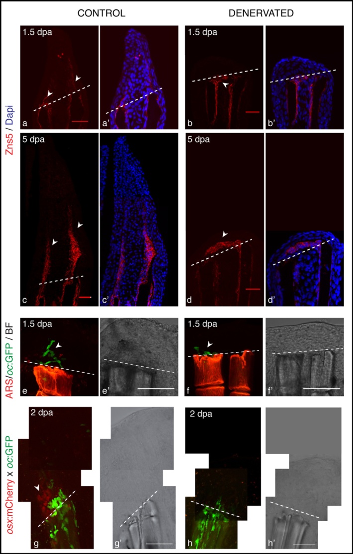
Scleroblasts alignment in amputated fins, upon denervation. a-d) Staining for Zns5 and DAPI in longitudinal sections shows that between 1 and 1.5 dpa, Zns5-positive cells start to accumulate just distal to the amputation plane in control fins (a-arrowhead). However, in denervated fins scleroblasts are not aligned with the stump rays, but instead are deposited between the 2 hemi-rays (b-arrowhead). At later time points the scleroblast deposition covers the tip of denervated rays (d). e,f) Live imaging with the Tg (oc:GFP) co-stained with Alizarin red-S (ARS). In vivo imaging of Tg (oc:GFP) stained with ARS shows that mature bone cells (oc-positive) of control fins are localized at the amputation level and in the blastema (e-arrowhead). However, in denervated fins oc-positive cells do not migrate further than the amputation plane (f-arrowhead). g,h) Live imaging with the transgenics Tg (oc:GFP), Tg (osx:mCherry) and ARS. Zebrafish transgenic line resulting from an outcross between Tg(oc:GFP) and Tg(osx:mCherry) shows that at 2 dpa only control fins present oc-positive and osx-positive cells in the blastema (g-arrowhead). a-h) The images are a projection of confocal optical slices. Dashed lines mark amputation plane. a-d) Scale bar - 25 µm. e-h) Scale bar - 100 µm.