
|
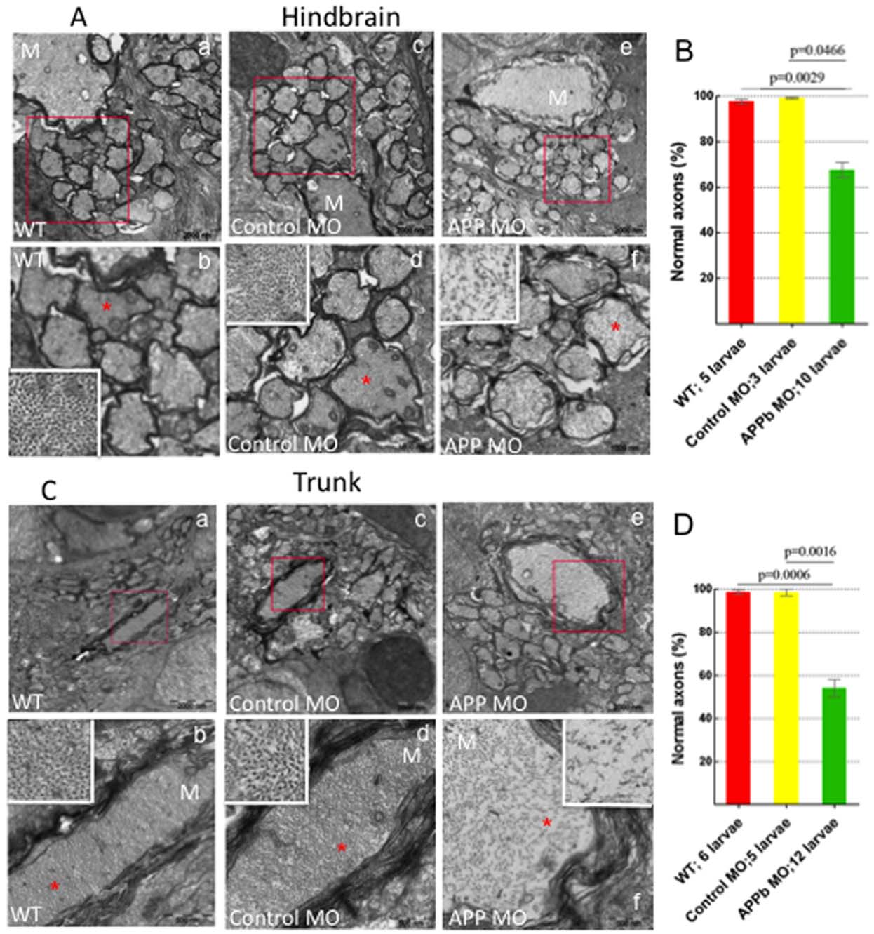
Fig. 4
Analysis of Transmission Electron Microscopy images of axons in zebrafish hindbrain and trunk regions (5 dpf).
(A) Transmission Electron Microscopy (TEM) images of axons in zebrafish hindbrain. Compared to uninjected and control MO-injected embryos, zebrafish embryos injected with APPb-MO (8 ng) expressed a decreased density and disorganization of the cytoskeleton in both the Mauthner (M) axons and the axons around the M axon. Panels b, d, and f are amplifications of the boxed areas in panels a, c and e, respectively. The white rectangles in panels b, d, and f are amplifications of the areas marked with red 5-point stars. (B) Quantifying the defects of axonal cytoskeletal morphology in the hindbrain of APPb morphant embryos. At 5 dpf, zebrafish embryos injected with 8 ng of APPb-MO showed a disruption in axon cytoskeletal dynamics in the hindbrain region. For the TEM experiment, the embryos were injected with 8 ng of the APPb MO instead of 10 ng. The embryos that were injected with 8 ng of the APPb MO experienced disorganization of the axonal cytoskeleton; the cytoskeleton of the embryos that were injected with 10 ng of APPb MO experienced severely defects in or loss of axons. Statistical significance was observed comparing uninjected (263 axons) and morphant (662 axons) larvae (p = 0.0029, p<0,05 in two-tailed paired t-test). Statistical significance was also observed between control MO (217 axons) and morphant larvae (p = 0.0466, p<0.05 in two tailed paired t-test). (C) TEM images of axons at in the zebrafish trunk section. Compared to uninjected and control MO-injected larvae, zebrafish larvae injected with 8 ng of APPb-MO expressed a decrease in axonal density and had defects in cytoskeletal organization of axons, including the M axon. Panels b, d, and f are amplifications of the boxed areas in panels a, c and e, respectively. The white rectangles in panels b, d and f are amplifications of the areas marked with red 5-point stars. (D) Quantifying defects of axonal cytoskeletal morphology in the trunk region of APPb morphant embryos. At 5 dpf, zebrafish embryos injected with 8 ng of APPb MO expressed an abnormal phenotype in axonal cytoskeletal organization in the trunk region. Statistical significance was observed comparing uninjected (217 axons) and morphant (562 axons) larvae (p = 0.0006, p<0.05 in two tailed paired t-test). Statistical significance was also observed between control MO (212 axons) and morphant larvae (p = 0.0016, p<0.05 in two tailed paired t-test).