
|
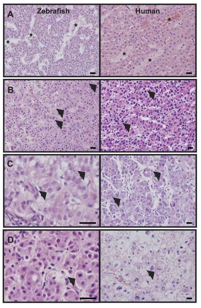
Fig. S2 Zebrafish livers with activated β-catenin share histologic features with human hepatocellular carcinoma (HCC).
(A) Adult zebrafish livers with activated β-catenin (left) and human HCC (right) show architectural disruption including enlarged interhepatic spaces resembling spongiosis hepatis (zebrafish) or peliosis hepatis (human) (asterisks). (B-D) Similarly, zebrafish (left) and human (right) samples show cytological abnormalities including nuclear enlargement and nuclear contour irregularities (arrows, B), prominent nucleoli (arrows, C), and mitotic figures (arrows, D). Hematoxylin and eosin (H&E) stained sections; scale bars, 20 µm.