
|
Fig. 8
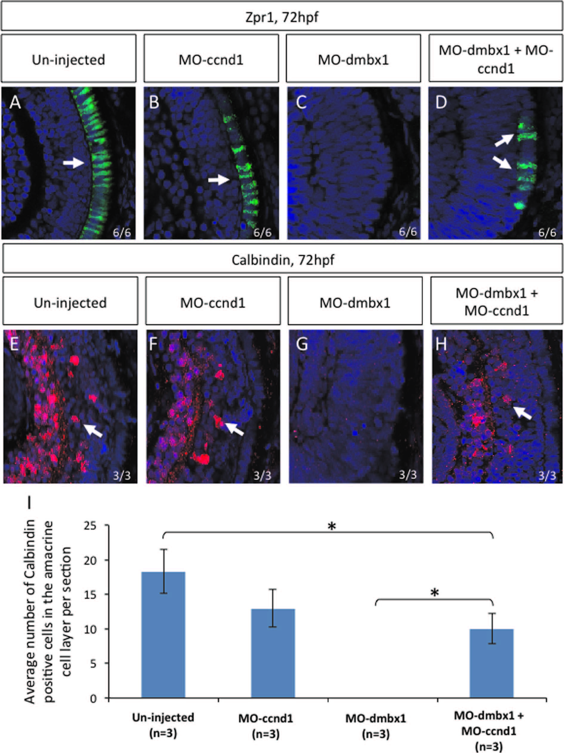
Repression of cyclinD1 partially rescued dmbx1 morphants phenotype. Coronal cryosections of 72 hpf retina (A–H). Retinal differentiation in the central retina can be seen using immunohistochemistry with Zpr1 (labels photoreceptors) and Calbindin (labels retinal ganglion+amacrine cells) markers. Both un-injected (A and E) and MOccnd1 (B and F) embryos express differentiation retinal markers. No differentiation markers are shown in dmbx1 morphants (MO1a+MO1b) (C and G). When dmbx1 morphants are knocked down with cyclin D1 morpholino (MO1a+MO1b+MOccnd1) (D and H), cells in the central retina are able to differentiate into specific cell types normally. Arrows point to the differentiation markers that are present in the retina. Numbers of Calbindin-positive cells observed in the amacrine cell layer per retinal section from each injection groups are summarized in graph (I), asterisks represent significant difference in the number of Calbindin-positive cells between two injection groups (p<0.05).
Reprinted from Developmental Biology, 402(2), Wong, L., Power, N., Miles, A., Tropepe, V., Mutual antagonism of the paired-type homeobox genes, vsx2 and dmbx1, regulates retinal progenitor cell cycle exit upstream of ccnd1 expression, 216-28, Copyright (2015) with permission from Elsevier. Full text @ Dev. Biol.