
|
Fig. 4
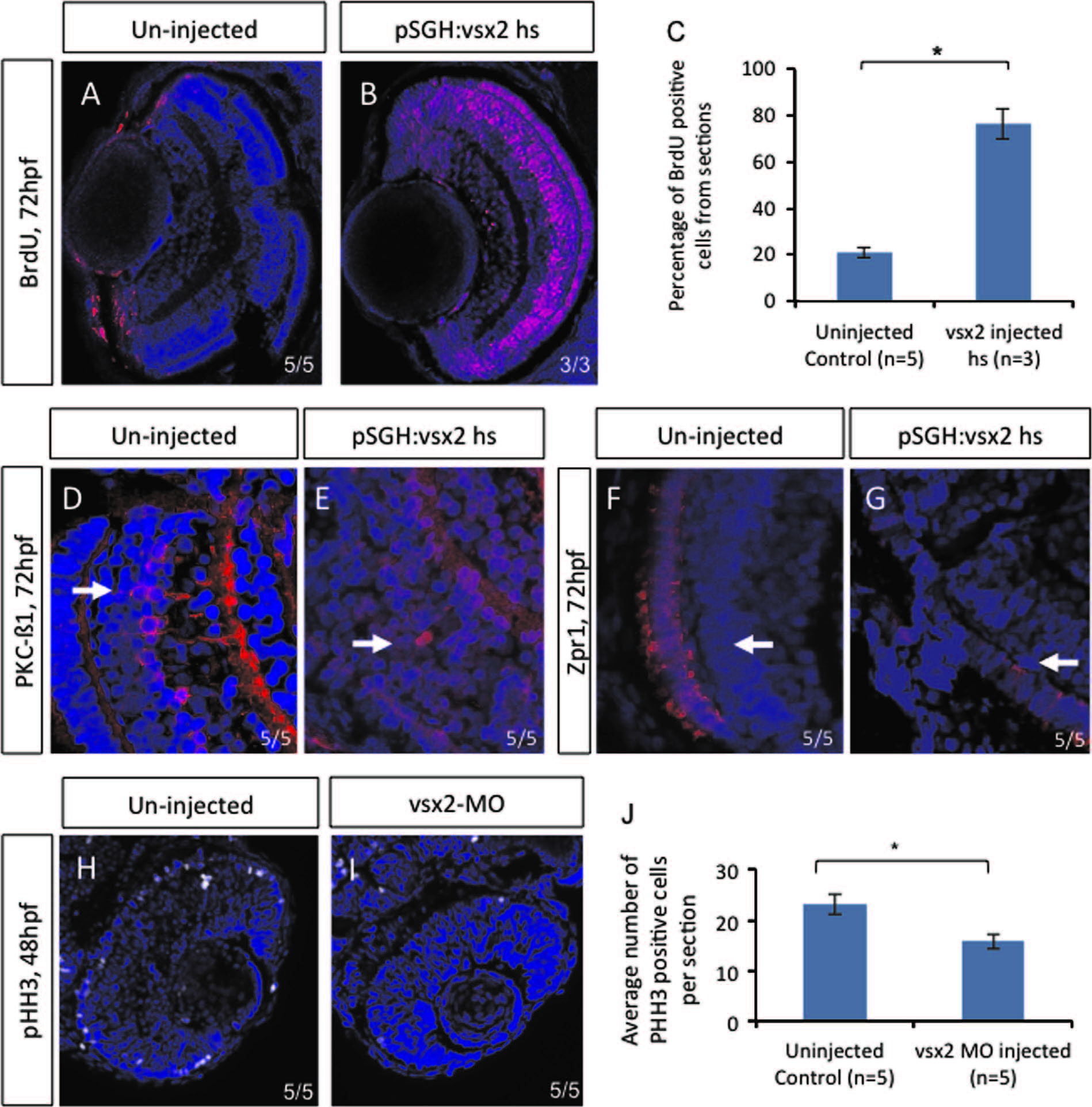
Over-expressing vsx2 embryos resulted in an increase of BrdU positive cells. Immunohistochemistry was performed on sections from uninjected (A) and pSGH:vsx2 injected (B) embryos that were heat shocked at 48 hpf and assessed at 72 hpf. There was a significant increase in the percentage of BrdU positive cells per section compared to uninjected (C). Standard deviation was calculated from four replicate experiments. These retinas also displayed delayed neuroretinal differentiation, as seen with PKC-B1 (D and E) and Zpr1 (F and G). Immunohistochemistry on sections from vsx2 morphants at 48 hpf showed a significant decrease in pHH3 cells (H–J). Statistical significance was assessed using Student′s t-test (p<0.05).
Reprinted from Developmental Biology, 402(2), Wong, L., Power, N., Miles, A., Tropepe, V., Mutual antagonism of the paired-type homeobox genes, vsx2 and dmbx1, regulates retinal progenitor cell cycle exit upstream of ccnd1 expression, 216-28, Copyright (2015) with permission from Elsevier. Full text @ Dev. Biol.