
|
Fig. 4
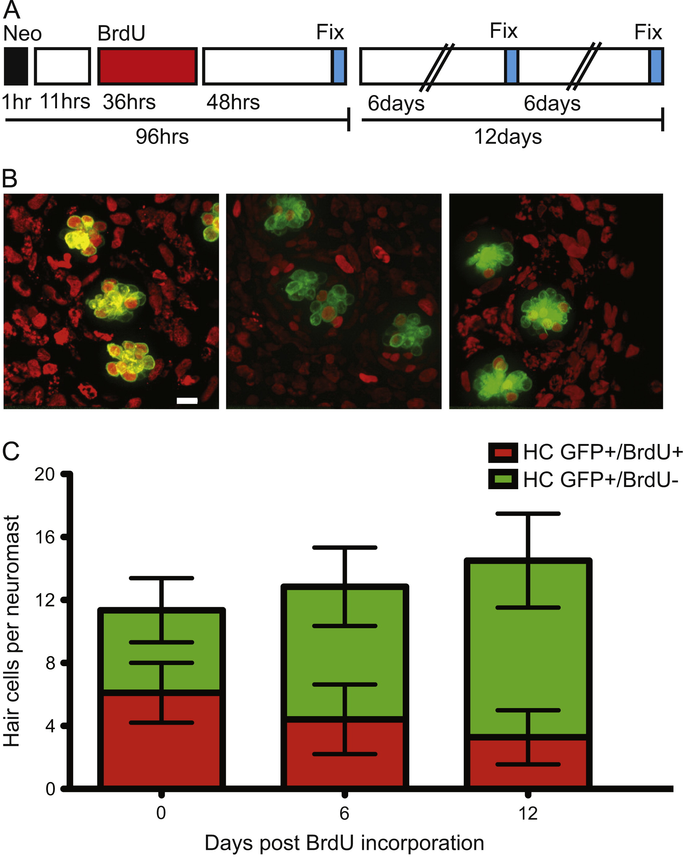
In adult zebrafish, BrdU labeled lateral line hair cells are lost over time under ambient conditions. (A) Schematic illustrating the experimental protocol: fish were treated with 400 µM neomycin for 1 h, then allowed to recover for 11 h, followed by a 36 h 5 mM BrdU incubation period, and finally fixed and stained at the indicated time-points. (B) Representative confocal z-stack maximum intensity projection images of neuromasts with hair cells labeled green with GFP and BrdU positive cell nuclei marked red. BrdU+ hair cells 0 days (left panel), 6 days (middle panel), and 12 days (right panel) after hair cell regeneration. Scale bar, 10 µM (C) Graph illustrating stacked averages (±s.d.) of GFP+ only hair cells (green) and GFP+ hair cells co-labeled with BrdU (red). N=2–3 adult zebrafish per condition, 30+ neuromasts per group.
Reprinted from Developmental Biology, 402(2), Cruz, I.A., Kappedal, R., Mackenzie, S.M., Hailey, D.W., Hoffman, T.L., Schilling, T.F., Raible, D.W., Robust regeneration of adult zebrafish lateral line hair cells reflects continued precursor pool maintenance, 229-38, Copyright (2015) with permission from Elsevier. Full text @ Dev. Biol.