
|
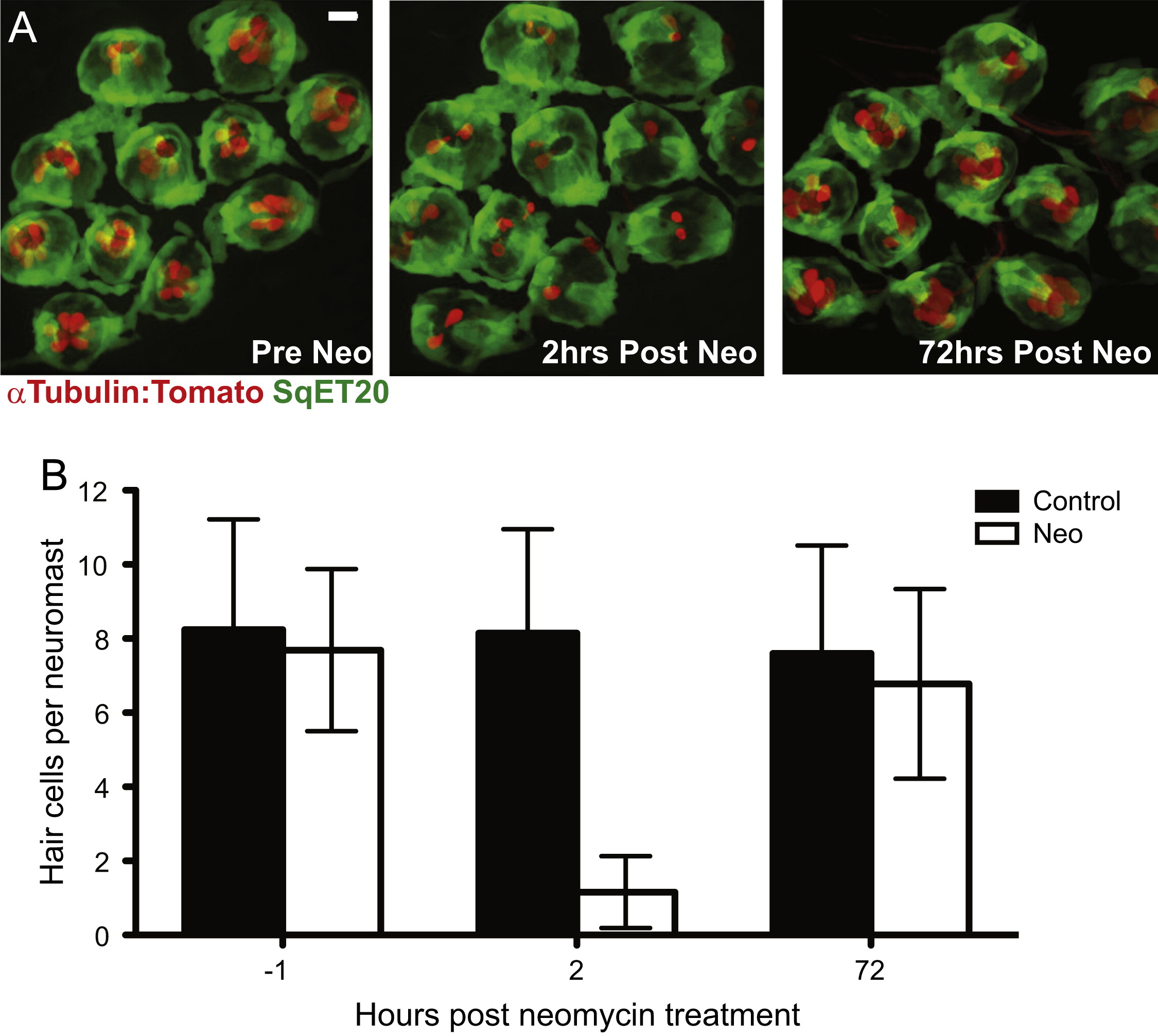
Fig. 1
Adult zebrafish lateral line hair cell regeneration after neomycin-induced ablation. Confocal z-stack maximum projection images of a peduncle stitch (11 neuromasts) of an adult sqet20Et;Tg(Ca-tuba1a:tdTomato) zebrafish with hair cells labeled with tdTomato (red) and a subset of support cells labeled with GFP (green). (A) Adult zebrafish peduncle neuromasts (left panel) were treated with 400 µM neomycin for 1 h (middle panel) and then allowed to recover for 72 h to assess hair cell regeneration (right panel). (B) Results are graphed as average number of hair cells per neuromast (±s.d.) for each treatment group. N=4 adult zebrafish per group, 40+ neuromasts per group. Scale bar, 10 µm.
Reprinted from Developmental Biology, 402(2), Cruz, I.A., Kappedal, R., Mackenzie, S.M., Hailey, D.W., Hoffman, T.L., Schilling, T.F., Raible, D.W., Robust regeneration of adult zebrafish lateral line hair cells reflects continued precursor pool maintenance, 229-38, Copyright (2015) with permission from Elsevier. Full text @ Dev. Biol.