
|
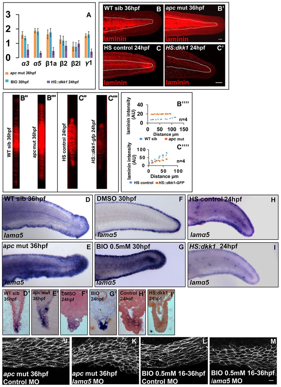
Fig. 7 Canonical Wnt signalling regulates the levels of laminins in the median fin fold. (A) Graph showing transcript level changes of laminins under gain and loss of canonical Wnt signalling by qRT-PCR. (B-C′′′′) Maximum intensity projections of laminin staining in the fin fold of sibling (B), apc mutant (B′), HS control (C) and HS::dkk-GFP (C2) embryos, their respective orthogonal sections (B",B′′′,C",C′′′) and plots comparing laminin intensities across PD axis in given genetic conditions (B′′′′,C′′′′). (D-I′) In situ hybridisation using lama5 probes in given genetic backgrounds or treatments (D-I) and sections of stained embryos (D′-I′). (J-M) Cell shape analysis in apc mutant (J,K) and 0.5 mM BIO-treated embryos (L,M) injected with control (J,L) or lama5 morpholino (K,M). The extent of cell stretching is reduced in the absence of lama5 function (K,M). Scale bars: 50 μm in B-C′ ; 10 μm in J-M.