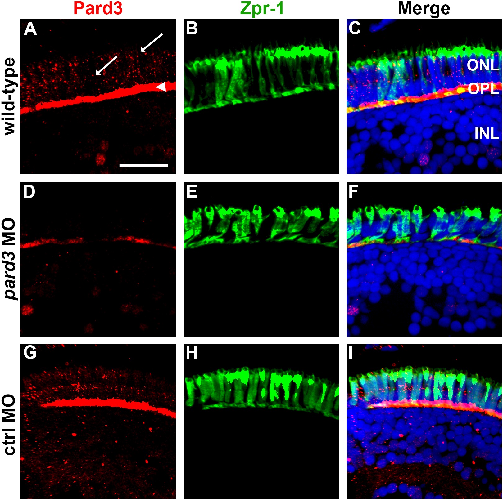
Fig. 2 Pard3 expression is reduced at 5 dpf following injection of pard3 morpholinos. (A–C) Transverse cryosections of wild-type retinas, (D–F) pard3 morphant retinas and (G–I) control morphant retinas with Pard3 antiserum (left column) and the monoclonal antibody zpr-1 (middle column). The right panels show the merged images. Pard3 immunoreactivity was seen in the outer plexiform layer (OPL) and at cell junctions (A, arrowhead and arrows, respectively) in wild-type and control MO retinas but was significantly reduced in morphants retinas. All sections were also counterstained with DAPI (blue). ONL = outer nuclear layer; OPL = outer plexiform layer; INL = inner nuclear layer. Scale bar = 10 μm.
                
                    
                        Image


                        Figure Caption


                        Figure Data


                        Acknowledgments


This image is the copyrighted work of the attributed author or publisher, and
ZFIN has permission only to display this image to its users.
Additional permissions should be obtained from the applicable author or publisher of the image.
      Full text @ PLoS One