
|
Fig. 1
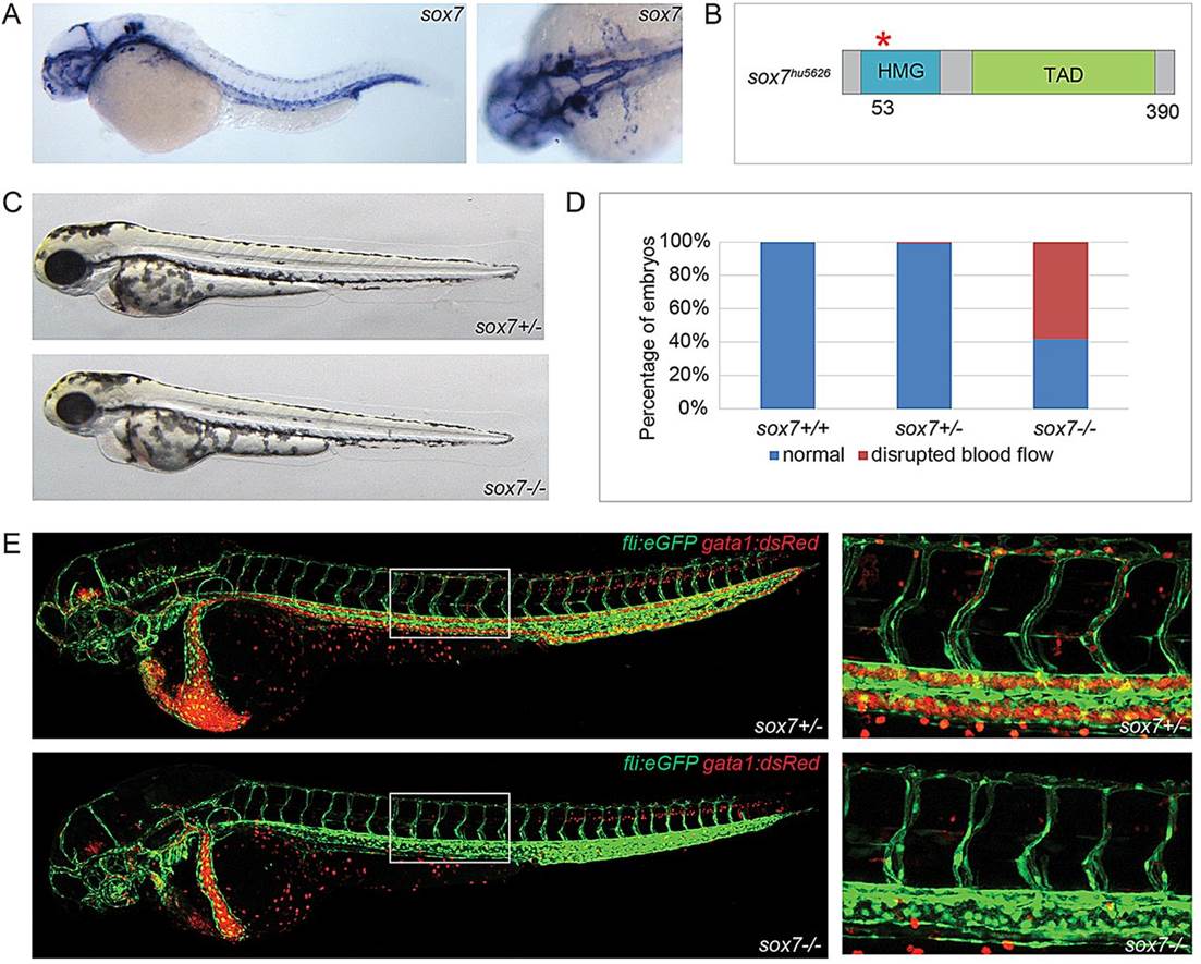
Disrupted blood circulation in sox7 zebrafish mutants. (A) sox7 in situ hybridization of 3dpf wild-type embryos (left panel, lateral view; right panel, dorsal view) showing sox7 expression in all main vessels. (B) Schematic diagram of the sox7hu5626 allele with a premature stop-codon after amino acid 53 (red asterisk). HMG, high mobility group box; TAD, trans-activating domain. (C) Overall normal appearance of sox7hu5626 heterozygous sibling and homozygous mutant at 2dpf. (D) On average, 59% of sox7hu5626 mutants display disturbed blood flow at 2.5dpf. Percentages of pooled embryos from four independent experiments (total of 275 embryos). Percentages can vary substantially between different backgrounds. (E) kdrl:GFP;gata1:dsRed-positive sox7hu5626 mutants lack functional blood circulation in the trunk, whereas heterozygous siblings have normal circulation at 2.5dpf. Right panels: higher magnifications of boxed area.