
|
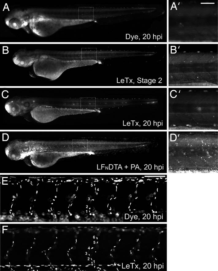
Fig. 3
LeTx does not induce generalized cell death or alter endothelial cell numbers in intact zebrafish embryonic vasculature. (A–D) Embryos were stained with acridine orange to examine cell death in dye-injected (A), LeTx-injected embryo at stage 2 or end stage of 20 hpi (B and C), or LFNDTA plus PA-injected (D) embryos. No significant difference was observed between dye and LeTx-injected embryos (A–C), whereas overall cell death was observed in those injected with LFNDTA plus PA (D) (each panel representative of n > 20 embryos). (A′–D′) Enlarged views of boxed regions. (E and F) Nuclei counts using the TG(fli1:nEGFP)y7 (20) line demonstrated equal endothelial cell numbers over the eight ISVs anterior to the cloacae between dye-injected controls (E), and embryos displaying a severe LeTx phenotype (F). There was no difference in the numbers of nuclei in this region between severe or mild phenotype and WT embryos (N = 3, n = 6 per condition; P = 0.378 and 0.887 respectively). Statistics were completed by using the Holm-Sidak method. (Scale bars, 80 µm.)