
|
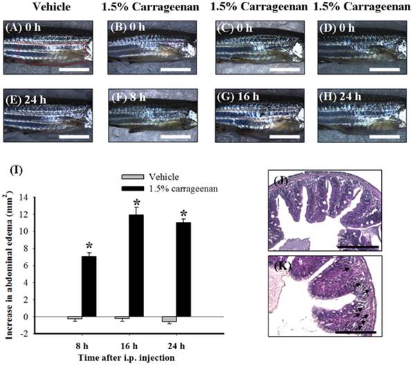
Fig. 2 The time course for the abdominal-edematous effects of i.p. carrageenan (1.5%) in adult zebrafish.
The photographic images show the gross pathology of the abdomen in the lateral view following i.p. injection of vehicle (PBS) plus i.p. injection vehicle (PBS) (A and E) and i.p. injection vehicle plus i.p. injection of 1.5% carrageenan at each time point (B–D and F–H). Images A–D were taken at 0 h after i.p. injection of vehicle or 1.5% carrageenan; images E, F, G, and H were taken at 24 h after i.p. injection of vehicle and 8, 16, and 24 h after 1.5% carrageenan injection. Scale bars: 5 mm for images A–H. The increase of the lateral area of the abdomen was given as a difference change from the basal values by subtracting the basal lateral area from the lateral area measured at each time point. Time course of change of the lateral area of abdomen induced by i.p. injection of vehicle (PBS) or carrageenan in adult zebrafish using a photographic image analysis system (I). Compared with the i.p. vehicle groups, the lateral area of abdomen increased progressively in the i.p. 1.5% carrageenan groups until 24 h. Sections (2 µm) of zebrafish abdominal tissues at 24 h after an i.p. injection of vehicle (J) or carrageenan (K). I.p. carrageenan dramatically induced leukocyte infiltration (arrows) in the intestine. Scale bars: 100 µm for images (J) and (K). Each bar in Figure (I) represents the mean ± SEM of 9 adult zebrafish per group. *P<0.05 compared with the same time points in the i.p. vehicle (PBS) group.